")
Nogo Antibody (C-4): sc-271878
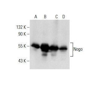
- Nogo Antibody (C-4) is a mouse monoclonal IgM κ Nogo antibody, cited in 27 publications, provided at 200 µg/ml
- specific for an epitope mapping between amino acids 18-41 at the N-terminus of Nogo of human origin
- recommended for detection of Nogo A, Nogo B and foocen of mouse, rat and human origin by WB, IP, IF, IHC(P) and ELISA; also reactive with additional species, including porcine
- available conjugated to agarose for IP; HRP for WB, IHC(P) and ELISA; phycoerythrin, FITC, Alexa Fluor® 488 or Alexa Fluor® 647 for IF, IHC(P) and FCM
- m-IgGκ BP-HRP is the preferred secondary detection reagent for Nogo Antibody (C-4) for WB and IHC(P) applications. This reagent is now offered in a bundle with Nogo Antibody (C-4) (see ordering information below). For additional m-IgGκ BP conjugates see our complete list of Mouse IgG Binding Proteins.

QUICK LINKS
SEE ALSO...
Nogo Antibody (C-4) is a mouse monoclonal IgM antibody that detects Nogo protein of mouse, rat, and human origin by western blotting (WB), immunoprecipitation (IP), immunofluorescence (IF), immunohistochemistry, and enzyme-linked immunosorbent assay (ELISA). anti-Nogo antibody (C-4) is available in both non-conjugated and various conjugated forms, including agarose, horseradish peroxidase (HRP), phycoerythrin (PE), fluorescein isothiocyanate (FITC), and multiple Alexa Fluor® conjugates. Nogo protein, also known as neurite outgrowth inhibitor or neuroendocrine secretion regulating protein, plays a critical role in the central nervous system by inhibiting axonal growth, which is essential for maintaining the integrity of CNS white matter. Nogo is primarily expressed by oligodendrocytes and is associated with the endoplasmic reticulum, where Nogo exists in three splice variants: Nogo-A, Nogo-B, and Nogo-C. Nogo presence in the CNS is crucial as Nogo prevents excessive axonal regeneration, which could lead to aberrant neural connections and potential dysfunction. Understanding Nogo interactions with other proteins, such as the NgR (Nogo receptor), is vital for elucidating Nogo′s role in neuroregeneration and the development of therapeutic strategies for spinal cord injuries and neurodegenerative diseases.
Alexa Fluor® is a trademark of Molecular Probes Inc., OR., USA
LI-COR® and Odyssey® are registered trademarks of LI-COR Biosciences
Nogo Antibody (C-4) References:
- Identification of the Nogo inhibitor of axon regeneration as a Reticulon protein. | GrandPré, T., et al. 2000. Nature. 403: 439-44. PMID: 10667797
- Function of Nogo-A/Nogo-A receptor in Alzheimer's disease. | Xu, YQ., et al. 2015. CNS Neurosci Ther. 21: 479-85. PMID: 25732725
- Nogo-A Antibodies for Progressive Multiple Sclerosis. | Ineichen, BV., et al. 2017. CNS Drugs. 31: 187-198. PMID: 28105588
- RTN4/NoGo-receptor binding to BAI adhesion-GPCRs regulates neuronal development. | Wang, J., et al. 2021. Cell. 184: 5869-5885.e25. PMID: 34758294
- RTN4/NoGo-receptor binding to BAI adhesion-GPCRs regulates neuronal development. | Wang, J., et al. 2022. Cell. 185: 218. PMID: 34995515
Ordering Information
| Product Name | Catalog # | UNIT | Price | Qty | FAVORITES | |
Nogo Antibody (C-4) | sc-271878 | 200 µg/ml | $322.00 | |||
Nogo Antibody (C-4): m-IgGκ BP-HRP Bundle | sc-522088 | 200 µg Ab, 40 µg BP | $361.00 | |||
Nogo Antibody (C-4) AC | sc-271878 AC | 500 µg/ml, 25% agarose | $424.00 | |||
Nogo Antibody (C-4) Alexa Fluor® 488 | sc-271878 AF488 | 200 µg/ml | $364.00 | |||
Nogo Antibody (C-4) Alexa Fluor® 647 | sc-271878 AF647 | 200 µg/ml | $364.00 | |||
Nogo Antibody (C-4) FITC | sc-271878 FITC | 200 µg/ml | $336.00 | |||
Nogo Antibody (C-4) HRP | sc-271878 HRP | 200 µg/ml | $322.00 | |||
Nogo (C-4) Neutralizing Peptide | sc-271878 P | 100 µg/0.5 ml | $69.00 | |||
Nogo Antibody (C-4) PE | sc-271878 PE | 200 µg/ml | $349.00 |