Date published: 2026-6-7

1-800-457-3801

SCBT Portrait Logo
Seach Input

Ep-CAM Antibody (VU-1D9): sc-51681

5.0(5)
Write a reviewAsk a question

Datasheets
  • Ep-CAM Antibody (VU-1D9) is a mouse monoclonal IgG1 κ, cited in 8 publications, provided at 200 µg/ml
  • raised against small cell lung carcinoma cell line HG9 of human origin
  • recommended for detection of Ep-CAM of human origin by WB, IP, IF, IHC(P) and FCM
  • available conjugated to either phycoerythrin or FITC for IF, IHC(P) and FCM
  • See Ep-CAM (C-10): sc-25308 for Ep-CAM antibody conjugates, including AC, HRP, FITC, PE, Alexa Fluor® 488, 594, 647, 680 and 790.
  • m-IgG Fc BP-HRP, m-IgG1 BP-HRP and m-IgGκ BP-HRP are the preferred secondary detection reagents for Ep-CAM Antibody (VU-1D9) for WB and IHC(P) applications. These reagents are now offered in bundles with Ep-CAM Antibody (VU-1D9) (see ordering information below).
Gene Editing Promo Banner

QUICK LINKS

SEE ALSO...

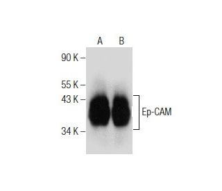
Ep-CAM Antibody (VU-1D9) is a mouse monoclonal IgG1 antibody that detects Ep-CAM in human samples through various applications including western blotting (WB), immunoprecipitation (IP), immunofluorescence (IF), immunohistochemistry with paraffin-embedded sections (IHCP), and flow cytometry (FCM). Ep-CAM, also known as tumor-associated calcium signal transducer 1 and MK-1, is a monomeric membrane glycoprotein predominantly expressed in normal human epithelial tissues and various carcinomas. The human Ep-CAM gene encodes a 314 amino acid protein that exists in two forms, with the major form being more prevalent and the minor form being reduced upon treatment with the amino-glycosylation inhibitor tunicamycin. Notably, Ep-CAM plays a crucial role in cell adhesion due to its extracellular domain, which contains two epidermal growth factor-like repeats followed by a cysteine-poor region essential for adhesive properties. This function is particularly important in maintaining tissue integrity and facilitating cellular communication, making Ep-CAM a significant marker for tumor progression and a potential target for therapeutic interventions in solid tumors. Anti-Ep-CAM antibody (VU-1D9) serves as a valuable tool for researchers aiming to explore Ep-CAM′s role in cancer biology and for developing targeted therapies.

For Research Use Only. Not Intended for Diagnostic or Therapeutic Use.

Ordering Information

Product NameCatalog #UNITPriceQtyFAVORITES

Ep-CAM Antibody (VU-1D9)

sc-51681
200 µg/ml
$322.00

Ep-CAM Antibody (VU-1D9): m-IgG Fc BP-HRP Bundle

sc-536850
200 µg Ab; 10 µg BP
$361.00

Ep-CAM Antibody (VU-1D9): m-IgGκ BP-HRP Bundle

sc-534017
200 µg Ab; 40 µg BP
$361.00

Ep-CAM Antibody (VU-1D9): m-IgG1 BP-HRP Bundle

sc-544857
200 µg Ab; 20 µg BP
$361.00

Ep-CAM Antibody (VU-1D9) FITC

sc-51681 FITC
200 µg/ml
$336.00

Ep-CAM Antibody (VU-1D9) PE

sc-51681 PE
200 µg/ml
$349.00

We are going to use this antibody first time with IHC -P application, which antigen retrivel can be used for this antibody (Acidic, citrate or alkaline, ph ? )please suggest Thank you

Asked by: Kaushal
Thank you for your question. You can find our recommended protocol with instructions on how to perform antigen unmasking under this link https://www.scbt.com/resources/protocols/immunoperoxidase-staining. We recommend using heat treatment with citrate buffer.
Answered by: Tech Support Europe
Date published: 2024-11-22

Can Ep-CAM (VU-1D9): sc-51681 monoclonal antibody be used in IF or IHC with mouse tissue or cells? Will there be non-specific staining?

Asked by: cjMara
Thank you for your inquiry. The use of mouse monoclonal antibodies with mouse samples is very common and typically poses no problem. To eliminate any potential cross-reactivity of an anti-mouse secondary detection reagent, Ep-CAM (VU-1D9): sc-51681 is available in a variety of direct conjugations, such as HRP, PE, FITC and AlexaFluors.
Answered by: Technical Support
Date published: 2017-02-24
  • y_2026, m_6, d_7, h_12CST
  • bvseo_bulk, prod_bvqa, vn_bulk_3.0.46
  • cp_1, bvpage1
  • co_hasquestionsanswers, tq_2
  • loc_en_US, sid_51681, prod, sort_[SortEntry(order=LAST_APPROVED_ANSWER_SUBMISSION_TIME, direction=DESCENDING)]
  • clientName_scbt
  • bvseo_sdk, java_sdk, bvseo-4.0.0
  • CLOUD, getContent, 179ms
  • QUESTIONS, PRODUCT
Rated 5 out of 5 by from Worked in WB in MCF7 and AWorked in WB in MCF7 and A-431 whole cell lysates. -SCBT QC
Date published: 2015-04-22
Rated 5 out of 5 by from Robust signal in WB in human transfectedRobust signal in WB in human transfected 293T whole cell lysates. -SCBT QC
Date published: 2015-04-15
Rated 5 out of 5 by from Positive signal in WB in human transfectedPositive signal in WB in human transfected 293T and A-431 whole cell lysates. -SCBT QC
Date published: 2015-04-14
Rated 5 out of 5 by from Good specificity in WB in MCF7 whole cell lysateGood specificity in WB in MCF7 whole cell lysate. -SCBT QC
Date published: 2015-04-10
Rated 5 out of 5 by from Beautiful immunoperoxidase membrane and cytoplasmicBeautiful immunoperoxidase membrane and cytoplasmic staining in formalin fixed, paraffin-embedded human rectum tissue. -SCBT QC
Date published: 2015-02-13
  • y_2026, m_6, d_7, h_12
  • bvseo_bulk, prod_bvrr, vn_bulk_3.0.46
  • cp_1, bvpage1
  • co_hasreviews, tv_0, tr_5
  • loc_en_US, sid_51681, prod, sort_[SortEntry(order=SUBMISSION_TIME, direction=DESCENDING)]
  • clientName_scbt
  • bvseo_sdk, java_sdk, bvseo-4.0.0
  • CLOUD, getReviews, 9ms
  • REVIEWS, PRODUCT
Ep-CAM Antibody (VU-1D9) is rated 5.0 out of 5 by 5.
  • y_2026, m_6, d_7, h_12
  • bvseo_bulk, prod_bvrr, vn_bulk_3.0.46
  • cp_1, bvpage1
  • co_hasreviews, tv_0, tr_5
  • loc_en_US, sid_51681, prod, sort_[SortEntry(order=SUBMISSION_TIME, direction=DESCENDING)]
  • clientName_scbt
  • bvseo_sdk, java_sdk, bvseo-4.0.0
  • CLOUD, getAggregateRating, 105ms
  • REVIEWS, PRODUCT