Date published: 2025-9-10

001 800-1338-3838

SCBT Portrait Logo
Seach Input

EDG-1/S1P1/S1PR1 항체 (A-6): sc-48356

4.0(13)
리뷰 쓰기질문하기

데이터 시트
  • EDG-1/S1P1/S1PR1 항체 A-6 는 마우스 monoclonal IgG3 κ EDG-1/S1P1/S1PR1 항체, 23간행물에 인용, 이며 200 µg/ml으로 제공합니다.
  • human origin의 EDG-1에서 내부에 위치한 322-381 아미노산을 항원으로 사용하였습니다.
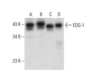
  • WB, IP, IF, IHC(P) 와ELISA으로 human origin의 EDG-1을 검출할것을 권장합니다.
  • IP에 사용가능한 agarose결합제품; HRP/b> for WB, IHC(P) and ELISA; IF, IHC(P) 와 FCM에 사용가능한 phycoerythrin/b>결합제품, FITC결합제품, Alexa Fluor® 488/b>결합제품 와 Alexa Fluor® 647/b> 결합제품 이 있습니다.
  • EDG-1/S1P1/S1PR1 (A-6): sc-48356무료10 µg 샘플을 신청하려면 기술지원팀 (또는 로컬 디스트리뷰터)에 연락주세요.
  • 현재 EDG-1/S1P1/S1PR1 항체(A-6)에 대해 선호하는 2차 검출 시약의 식별을 아직 완료하지 못했습니다. 이 작업은 진행 중입니다.

빠른 링크

더보기

EDG-1 항체(A-6)는 웨스턴 블롯팅(WB), 면역 침전(IP), 면역 형광(IF), 면역 조직 화학 및 효소 결합 면역 흡착 분석(ELISA)으로 인간 유래의 EDG-1을 검출하는 마우스 단일 클론 IgG3 카파 경쇄 항체입니다. EDG-1(A-6) 항체는 비접합 형태와 아가로스, 홀세라디시 퍼옥시다제(HRP), 피코에리스린(PE), 플루오레세인 이소티오시안산염(FITC), 다중 Alexa Fluor® 접합체를 포함한 다양한 접합 형태 모두에서 사용할 수 있습니다. S1PR1 또는 S1P1로도 알려진 EDG-1은 스핑고신-1-인산염(S1P) 및 리소포스파티딘산(LPA)과 같은 리소인지질 신호 분자의 효과를 매개하는 데 중요한 역할을 하는 G 단백질 결합 수용체의 내피 분화 유전자(EDG) 계열의 일원입니다. EDG-1은 주로 세포 표면에 위치하며 Gi 단백질과 결합하여 Akt와 같은 하류 신호 전달 경로를 활성화함으로써 세포 생존, 성장, 이동, 분화 등 다양한 세포 과정에 관여합니다. 이는 특히 신경교종 세포에서 중요한데, EDG-1 발현은 종양 진행 및 생존과 관련이 있습니다. 여러 G 단백질과 상호 작용하는 EDG-1의 능력은 광범위한 생리적 반응에 영향을 미치기 때문에 암 생물학 및 치료제 개발 연구의 중요한 표적이 되고 있습니다.

연구용으로만 사용가능합니다. 진단이나 치료용으로 사용불가합니다.

Alexa Fluor®는 미국 오리건주 Molecular Probes Inc.의 상표입니다.

LI-COR®와 Odyssey®는 LI-COR Biosciences의 등록 상표입니다.

EDG-1/S1P1/S1PR1 항체 (A-6) 참고문헌:

  1. 스핑고신 1-인산 수용체 EDG-1, EDG-3, EDG-5의 차별적 약리학적 특성과 신호 전달.  |  Ancellin, N. and Hla, T. 1999. J Biol Chem. 274: 18997-9002. PMID: 10383399
  2. 리소인지질과 리소스핑고지질에 대한 G 단백질 결합 세포 수용체의 하위 계열.  |  Goetzl, EJ. and An, S. 1999. Adv Exp Med Biol. 469: 259-64. PMID: 10667339
  3. 스핑고신-1-포스페이트는 G 단백질 결합 수용체 EDG-6의 리간드입니다.  |  Van Brocklyn, JR., et al. 2000. Blood. 95: 2624-9. PMID: 10753843
  4. EDG-1 계열의 G-단백질 결합 수용체를 통한 스핑고신-1-인산 신호 전달.  |  Hla, T., et al. 2000. Ann N Y Acad Sci. 905: 16-24. PMID: 10818438
  5. 스핑고신-1-인산: EDG-1 계열의 G-단백질 결합 수용체의 리간드.  |  Spiegel, S. 2000. Ann N Y Acad Sci. 905: 54-60. PMID: 10818441
  6. 포유류 세포의 스핑고신 1-인산염 신호.  |  Pyne, S. and Pyne, NJ. 2000. Biochem J. 349: 385-402. PMID: 10880336
  7. C6 신경교종 세포의 신호 전달 경로에서 스핑고신 1-인산 수용체인 Edg-1과 Edg-5의 차별적 역할.  |  Sato, K., et al. 2000. Brain Res Mol Brain Res. 85: 151-60. PMID: 11146117
  8. 매트리겔 모델 기저막을 통한 유르캇 T 세포 이동에 대한 리소포스파티딘산 수용체 선택적 효과.  |  Zheng, Y., et al. 2001. J Immunol. 166: 2317-22. PMID: 11160288
  9. 성체 쥐 뇌의 Edg2 수용체 분포.  |  Handford, EJ., et al. 2001. Neuroreport. 12: 757-60. PMID: 11277579
  10. 스핑고신 1-인산염은 내피 세포에서 Gi 단백질/포스포이노시타이드 3-키나아제 경로를 통해 Akt, 산화질소 생성 및 화학 주성을 활성화합니다.  |  Morales-Ruiz, M., et al. 2001. J Biol Chem. 276: 19672-7. PMID: 11278592
  11. 스핑고신 1-인산 자극 세포에서 Edg-1/S1P1의 역학에 있어서 N-글리코실화 작용의 역할.  |  Kohno, T. and Igarashi, Y. 2004. Glycoconj J. 21: 497-501. PMID: 15750791

주문정보

제품명카탈로그 번호 단위가격수량관심품목

EDG-1/S1P1/S1PR1 항체 (A-6)

sc-48356
200 µg/ml
$316.00

EDG-1/S1P1/S1PR1 항체 (A-6) AC

sc-48356 AC
500 µg/ml, 25% agarose
$416.00

EDG-1/S1P1/S1PR1 항체 (A-6) Alexa Fluor® 488

sc-48356 AF488
200 µg/ml
$357.00

EDG-1/S1P1/S1PR1 항체 (A-6) Alexa Fluor® 647

sc-48356 AF647
200 µg/ml
$357.00

EDG-1/S1P1/S1PR1 항체 (A-6) FITC

sc-48356 FITC
200 µg/ml
$330.00

EDG-1/S1P1/S1PR1 항체 (A-6) HRP

sc-48356 HRP
200 µg/ml
$316.00

EDG-1/S1P1/S1PR1 항체 (A-6) PE

sc-48356 PE
200 µg/ml
$343.00

If I Stored the antibody at -20° C, does it affect its activity?

Asked by: June 0615
Thank you for your question. EDG-1/S1P1/S1PR1 抗体 (A-6): sc-48356 should be stored at 4° C, do not freeze. If it is stored at -20° C, it may affect its activity.
Answered by: Technical Service SH
Date published: 2022-02-28

What is the recommended secondary detection reagent to be used with the EDG-1 Antibody (A-6): sc-48356 for Western blot?

Asked by: Trav11
Thank you for your question. We recommend using one of our exclusive Mouse IgG Binding Proteins as a secondary detection reagent, such as m-IgGκ BP-HRP: sc-516102, for Western blot. A complete list of all available binding proteins is available on our website here: https://www.scbt.com/scbt/browse/support-products-mouse-igg-binding-proteins/_/N-ecrety If you have any further questions or concerns, please feel free to contact our Technical Service department by calling 800-457-8301 option 2, emailing scbt@scbt.com, or using the Live Chat function on our website.
Answered by: Technical Support 15
Date published: 2017-01-22
  • y_2025, m_9, d_9, h_6CST
  • bvseo_bulk, prod_bvqa, vn_bulk_3.0.42
  • cp_1, bvpage1
  • co_hasquestionsanswers, tq_2
  • loc_ko_KR, sid_48356, prod, sort_[SortEntry(order=LAST_APPROVED_ANSWER_SUBMISSION_TIME, direction=DESCENDING)]
  • clientName_scbt
  • bvseo_sdk, java_sdk, bvseo-4.0.0
  • CLOUD, getContent, 115ms
  • QUESTIONS, PRODUCT
Rated 4 out of 5 by from Works wellDoesn't seem to give a super robust signal, but it does work. We like to try and re-use antibodies for Westerns, however this antibody seems to only work when fresh, at a 1:250 dilution.
Date published: 2019-01-14
Rated 5 out of 5 by from worked well1:50 dilution for IF with human brain section, worked well ,expected staining.
Date published: 2018-05-26
Rated 5 out of 5 by from Working very good for ImmunoblottingI bought a free sample of this antibody. It is working excellent. Clean band.
Date published: 2017-09-18
Rated 5 out of 5 by from Detects GFP tagged and endogenous EDG-1 in IFAntibody detects successfully GFP tagged and endogenous EDG-1 in human cell lines.
Date published: 2017-08-30
Rated 1 out of 5 by from Did not work in mouse tissueI received a free sample of this because H-60 has been discontinued. I tested in both IHC and Western. There was no staining in IHC and the Western showed no bands. I have attached a blot from the H-60 antibody that I have that is >4 years old. I was really hoping the monoclonal would work. I used the same tissue and protocol for both Westerns. Santa Cruz person I worked with was great, no complaints there.
Date published: 2017-07-05
Rated 1 out of 5 by from Not good for immunohistochemistryWe obtained a sample of this antibody to test since the EDG-1 (H-60) is no longer available. The signal was not above the background signal and it did not label any structures in the cell specifically when compared to our old antibody.
Date published: 2017-06-15
Rated 5 out of 5 by from Great productVery nice western blot data, strong, clear signal; also strong IHC staining results
Date published: 2017-02-02
Rated 5 out of 5 by from Excellent!This antibody works great in western blot analysis of EDG-1 expression in human brain tissue extract.
Date published: 2017-01-11
  • y_2025, m_9, d_9, h_6
  • bvseo_bulk, prod_bvrr, vn_bulk_3.0.42
  • cp_1, bvpage1
  • co_hasreviews, tv_0, tr_13
  • loc_ko_KR, sid_48356, prod, sort_[SortEntry(order=SUBMISSION_TIME, direction=DESCENDING)]
  • clientName_scbt
  • bvseo_sdk, java_sdk, bvseo-4.0.0
  • CLOUD, getReviews, 9ms
  • REVIEWS, PRODUCT
EDG-1/S1P1/S1PR1 항체 (A-6) is rated 4.0 out of 5 by 13.
  • y_2025, m_9, d_9, h_6
  • bvseo_bulk, prod_bvrr, vn_bulk_3.0.42
  • cp_1, bvpage1
  • co_hasreviews, tv_0, tr_13
  • loc_ko_KR, sid_48356, prod, sort_[SortEntry(order=SUBMISSION_TIME, direction=DESCENDING)]
  • clientName_scbt
  • bvseo_sdk, java_sdk, bvseo-4.0.0
  • CLOUD, getAggregateRating, 120ms
  • REVIEWS, PRODUCT