
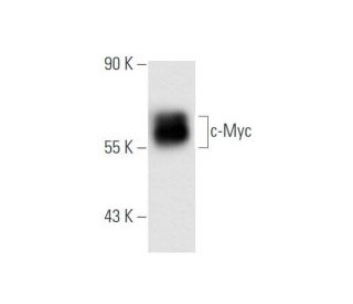
Myc Antibody (1.N.2): sc-70469
- Myc Antibody (1.N.2) is a mouse monoclonal IgG1 κ, cited in 16 publications, provided at 200 µg/ml
- raised against full length c-Myc of human origin
- recommended for detection of c-Myc p67 and c-Myc tagged fusion proteins of mouse, rat, human and avian origin by WB, IP and IF
- See Myc (9E10): sc-40 for Myc antibody conjugates, including AC, HRP, FITC, PE, Alexa Fluor® 488, 594, 647, 680 and 790.
- m-IgGκ BP-HRP is the preferred secondary detection reagent for Myc Antibody (1.N.2) for WB applications. This reagent is now offered in a bundle with Myc Antibody (1.N.2) (see ordering information below). For additional m-IgGκ BP conjugates see our complete list of Mouse IgG Binding Proteins.

c-Myc Antibody (1.N.2) is a mouse monoclonal IgG1 kappa light chain antibody raised against full-length human c-Myc, a vital transcription factor involved in regulating numerous cellular processes, including cell proliferation, metabolism, and apoptosis. c-Myc mouse monoclonal antibody (1.N.2) recognizes an epitope within the c-Myc immunogen sequence. Anti-c-Myc monoclonal antibody (1.N.2) has proven crucial for advancing research on c-Myc′s role in oncogenic pathways and the development of therapeutic strategies for c-Myc-driven cancers. The c-Myc protein, encoded by the MYC gene, belongs to the Myc family along with N-Myc and L-Myc. c-Myc exerts influence on gene expression by binding to specific E-box sequences in DNA, regulating numerous genes involved in cell cycle control and metabolic pathways. The activity of c-Myc depends on heterodimerization with Max protein, facilitated by the basic helix-loop-helix leucine zipper (bHLH-Zip) domain. The c-Myc/Max complex binds to DNA in a sequence-specific manner, driving the transcription of genes necessary for the G1 to S phase transition, as well as genes regulating cellular growth, metabolism, and mitochondrial function. This transcriptional control maintains normal cellular homeostasis, but overexpression or dysregulation of c-Myc frequently occurs in cancers such as lung, breast, and colon carcinomas, contributing to unchecked cell proliferation, resistance to apoptosis, and tumor progression. Regarding subcellular localization, c-Myc predominantly resides in the nucleus, where c-Myc functions as a transcriptional regulator. Nuclear localization proves crucial for c-Myc′s ability to interact with DNA and orchestrate the transcriptional programs controlling cell growth and division. c-Myc can shuttle between the nucleus and cytoplasm, a dynamic localization important for regulation. Under certain conditions, c-Myc may undergo sequestration in the cytoplasm, altering availability for DNA binding and transcriptional activity. This ability to transition between cellular compartments allows for tight regulation of c-Myc′s role in cellular function and transformation.
Alexa Fluor® is a trademark of Molecular Probes Inc., OR., USA
LI-COR® and Odyssey® are registered trademarks of LI-COR Biosciences
Myc Antibody (1.N.2) References:
- c-Myc-regulated microRNAs modulate E2F1 expression. | O'Donnell, KA., et al. 2005. Nature. 435: 839-43. PMID: 15944709
- Crosstalk between c-Myc and ribosome in ribosomal biogenesis and cancer. | Dai, MS. and Lu, H. 2008. J Cell Biochem. 105: 670-7. PMID: 18773413
- Nucleophosmin is essential for c-Myc nucleolar localization and c-Myc-mediated rDNA transcription. | Li, Z. and Hann, SR. 2013. Oncogene. 32: 1988-94. PMID: 22665062
- c-Myc modulates microRNA processing via the transcriptional regulation of Drosha. | Wang, X., et al. 2013. Sci Rep. 3: 1942. PMID: 23735886
- Fluorescent Dansyl-Guanosine Conjugates that Bind c-MYC Promoter G-Quadruplex and Downregulate c-MYC Expression. | Pavan Kumar, Y., et al. 2016. Chembiochem. 17: 388-93. PMID: 26708259
- The deubiquitinase USP38 promotes cell proliferation through stabilizing c-Myc. | Xu, Z., et al. 2021. Int J Biochem Cell Biol. 137: 106023. PMID: 34102342
- HELZ2 promotes K63-linked polyubiquitination of c-Myc to induce retinoblastoma tumorigenesis. | Dai, H., et al. 2021. Med Oncol. 39: 11. PMID: 34761308
- Identification of Trovafloxacin, Ozanimod, and Ozenoxacin as Potent c-Myc G-Quadruplex Stabilizers to Suppress c-Myc Transcription and Myeloma Growth. | Zhang, J., et al. 2022. Mol Inform. 41: e2200011. PMID: 35355429
- Structure-based discovery of Licoflavone B and Ginkgetin targeting c-Myc G-quadruplex to suppress c-Myc transcription and myeloma growth. | Liu, L., et al. 2022. Chem Biol Drug Des. 100: 525-533. PMID: 35557489
- FIT links c-Myc and P53 acetylation by recruiting RBBP7 during colorectal carcinogenesis. | Guo, L., et al. 2023. Cancer Gene Ther. 30: 1124-1133. PMID: 37225855
Ordering Information
| Product Name | Catalog # | UNIT | Price | Qty | FAVORITES | |
Myc Antibody (1.N.2) | sc-70469 | 200 µg/ml | $322.00 | |||
Myc Antibody (1.N.2): m-IgGκ BP-HRP Bundle | sc-552808 | 200 µg Ab; 40 µg BP | $361.00 |