")
TBK1 Antikörper (A-6): sc-398366
- TBK1 Antikörper A-6 ist ein Maus monoklonales IgG1 κ TBK1 Antikörper, verwendet in 12 wissenschaftlichen Veröffentlichungen, in einer Menge von 200 µg/ml
- gezogen gegen die Aminosäuresequenz 355-729 lokalisiert am C-terminus von TBK1 aus der Spezies mouse
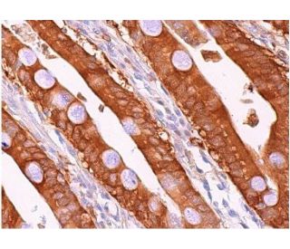
- TBK1 Antikörper (A-6) ist empfohlen für die Detektion von TBK1 aus der Spezies mouse, rat und human per WB, IP, IF, IHC(P) und ELISA
- Anti-TBK1 Antikörper (A-6) ist erhältlich als Konjugat mit Agarose für IP; HRP für WB, IHC(P) und ELISA; und entweder mit Phycoerythrin oder FITC für IF, IHC(P) und FCM
- auch erhältlich als Konjugat mit Alexa Fluor® 488, Alexa Fluor® 546, Alexa Fluor® 594 oder Alexa Fluor® 647 für IF, IHC(P) und FCM
- auch erhältlich als Konjugat mit Alexa Fluor® 680 oder Alexa Fluor® 790 für WB (NIR), IF und FCM
- m-IgG Fc BP-HRP, 1 BP-HRP">m-IgG1 BP-HRP und m-IgGκ BP-HRP sind die bevorzugten sekundären Nachweisreagenzien für TBK1 Antikörper (A-6) für WB- und IHC(P)-Anwendungen. Diese Reagenzien werden jetzt in Bündeln mit TBK1 Antikörper (A-6) angeboten(siehe Bestellinformationen unten).

Direktverknüpfungen
Siehe auch...
Der TBK1-Antikörper (A-6) ist ein monoklonaler Maus IgG1 κ TBK1-Antikörper (auch als NAK-Antikörper, T2K-Antikörper, Encephalopathie, akut, infektionsbedingt, herpes-spezifisch, 8 (IIAE8) Antikörper oder Frontotemporale Demenz und/oder Amyotrophe Lateralsklerose 4 (FTDALS4) Antikörper bezeichnet), der das TBK1-Protein von Maus-, Ratte- und menschlicher Herkunft mittels WB, IP, IF, IHC(P) und ELISA detektiert. Der TBK1-Antikörper (A-6) ist sowohl in nicht konjugierter Form als auch in mehreren konjugierten Formen des TBK1-Antikörpers erhältlich, darunter Agarose, HRP, PE, FITC und mehrere Alexa Fluor®-Konjugate. Der Transkriptionsfaktor NFkB wird im Zytoplasma in einer inaktiven Form durch das inhibitory Protein IkB gehalten. Die Aktivierung von NFkB erfordert, dass IkB an bestimmten Serinresten phosphoryliert wird, was zu einer gezielten Degradation von IkB führt. IkB-Kinase α (IKKα), früher als CHUK bezeichnet, interagiert mit IkB-a und phosphoryliert spezifisch IkB-α an den Stellen, die seine Degradation auslösen, Serin 32 und 36. Das funktionelle IKK-Komplex enthält drei Untereinheiten, IKKα, IKKβ und IKKγ (auch als NEMO bezeichnet), und jede scheint einen essentiellen Beitrag zur Phosphorylierung von IkB zu leisten. Die TANK-Bindungskinase (TBK1), auch als T2K bezeichnet, ist eine neuartige IKK-verwandte Kinase, die in murinen und menschlichen Geweben identifiziert wurde. TBK1 wurde gezeigt, dass es sich mit TRAF2 und TANK in der NFkB-Aktivierungspathway komplexiert. TBK1 weist eine Homologie mit IKKα und IKKβ im Amino-Terminalteil auf, der die Kinase-Domäne einschließt.
Alexa Fluor® ist ein Markenzeichen von Molecular Probes Inc., OR., USA
LI-COR® und Odyssey® sind Markenzeichen von LI-COR Biosciences
TBK1 Antikörper (A-6) Literaturhinweise:
- STING spezifiziert die IRF3-Phosphorylierung durch TBK1 im zytosolischen DNA-Signalweg. | Tanaka, Y. and Chen, ZJ. 2012. Sci Signal. 5: ra20. PMID: 22394562
- TRIM23 vermittelt die virusinduzierte Autophagie über die Aktivierung von TBK1. | Sparrer, KMJ., et al. 2017. Nat Microbiol. 2: 1543-1557. PMID: 28871090
- Raum-zeitliche Kontrolle der ULK1-Aktivierung durch NDP52 und TBK1 während der selektiven Autophagie. | Vargas, JNS., et al. 2019. Mol Cell. 74: 347-362.e6. PMID: 30853401
- Die Rekrutierung von TBK1 zu STING aktiviert sowohl IRF3 als auch NF-κB, die die Immunabwehr gegen Tumore und Virusinfektionen vermitteln. | Yum, S., et al. 2021. Proc Natl Acad Sci U S A. 118: PMID: 33785602
- Tilirosid zielt auf TBK1, um Ferroptose zu induzieren und hepatozelluläre Karzinome für Sorafenib zu sensibilisieren. | Yang, C., et al. 2023. Phytomedicine. 111: 154668. PMID: 36657316
- VANGL2 hemmt die antivirale IFN-I-Signalisierung, indem es TBK1 für den autophagischen Abbau anvisiert. | Hu, Z., et al. 2023. Sci Adv. 9: eadg2339. PMID: 37352355
- ATG4B hemmt die antivirale Immunität durch GABARAP-gesteuerten autophagischen Abbau von TBK1. | Xie, W., et al. 2023. Autophagy. 19: 2853-2868. PMID: 37434364
- Regulierung des Transferrinrezeptor-Transports durch Optineurin und seine krankheitsassoziierten Mutanten. | Yamano, K., et al. 2024. EMBO J. 43: 754-779. PMID: 38287189
- Eine TBK1-Variante verursacht bei Mäusen Autophagolysosomal- und Motoneuronpathologie ohne Neuroinflammation. | Brenner, D., et al. 2024. J Exp Med. 221: PMID: 38517332
- CDK4/6 verändert die TBK1-Phosphorylierung, um den STING-Signalweg bei Prostatakrebs zu hemmen. | Li, W., et al. 2024. Cancer Res. 84: 2588-2606. PMID: 38861362
Bestellinformation
| Produkt | Katalog # | EINHEIT | Preis | ANZAHL | Favoriten | |
TBK1 Antikörper (A-6) | sc-398366 | 200 µg/ml | $322.00 | |||
TBK1 (A-6): m-IgG Fc BP-HRP Bundle | sc-530775 | 200 µg Ab; 10 µg BP | $361.00 | |||
TBK1 (A-6): m-IgGκ BP-HRP Bundle | sc-524312 | 200 µg Ab, 40 µg BP | $361.00 | |||
TBK1 (A-6): m-IgG1 BP-HRP Bundle | sc-544247 | 200 µg Ab; 20 µg BP | $361.00 | |||
TBK1 Antikörper (A-6) AC | sc-398366 AC | 500 µg/ml, 25% agarose | $424.00 | |||
TBK1 Antikörper (A-6) HRP | sc-398366 HRP | 200 µg/ml | $322.00 | |||
TBK1 Antikörper (A-6) FITC | sc-398366 FITC | 200 µg/ml | $336.00 | |||
TBK1 Antikörper (A-6) PE | sc-398366 PE | 200 µg/ml | $349.00 | |||
TBK1 Antikörper (A-6) Alexa Fluor® 488 | sc-398366 AF488 | 200 µg/ml | $364.00 | |||
TBK1 Antikörper (A-6) Alexa Fluor® 546 | sc-398366 AF546 | 200 µg/ml | $364.00 | |||
TBK1 Antikörper (A-6) Alexa Fluor® 594 | sc-398366 AF594 | 200 µg/ml | $364.00 | |||
TBK1 Antikörper (A-6) Alexa Fluor® 647 | sc-398366 AF647 | 200 µg/ml | $364.00 | |||
TBK1 Antikörper (A-6) Alexa Fluor® 680 | sc-398366 AF680 | 200 µg/ml | $364.00 | |||
TBK1 Antikörper (A-6) Alexa Fluor® 790 | sc-398366 AF790 | 200 µg/ml | $364.00 |