")
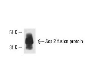
: sc-55478. Western Blot Analyse des humanen rekombinanten Sos 2-Fusionsproteins.")
Sos 2 Antikörper (A-8): sc-55478
- Sos 2 Antikörper (A-8) ist ein Maus monoklonales IgG2a (kappa light chain) in einer Menge von 200 µg/ml
- gezogen gegen Aminosäuren 1091-1170 gelegen in der Nähe vom C-terminus von Sos 2 aus der Spezies human
- Empfohlen für die Detektion von Sos 2 aus der Spezies human per WB, IP, IF und ELISA
- m-IgG Fc BP-HRP ist das bevorzugte sekundäre Nachweisreagenz für Sos 2 Antikörper (A-8) für WB-Anwendungen. Dieses Reagenz wird jetzt in einem Paket mit Sos 2 Antikörper (A-8) angeboten(siehe Bestellinformationen unten).

Direktverknüpfungen
Die Superfamilie der GTP-bindenden Proteine, zu denen die Ras-Proteine Prototypen sind, wurde in einer breiten Palette biologischer Aktivitäten impliziert. Studien haben eine Familie guaniner Nukleotid-freisetzender Faktoren (GRFs) identifiziert, die Ras in Säugetierzellen aktivieren, und ein "Adapter"-Protein (Sem 5/GRB2), das die Interaktion von GRFs mit aktivierten Rezeptormolekülen zu vermitteln scheint. Ras-GRF p140 fördert den Nukleotidaustausch an Ras p21s, aber nicht an anderen Mitgliedern der Ras-Gensuperfamilie. Darüber hinaus wurden drei Säugetierhomologe des Drosophila Ras-GRF, Son of Sevenless (Sos), beschrieben. Dazu gehören zwei aus der Maus, mSos 1 und mSos 2, und einer aus dem Menschen, hSos. Vav p95 wurde berichtet, als GRF bei der Aktivierung von Ras durch den T-Zell-Rezeptor zu funktionieren und wurde berichtet, eine Domäne ähnlich der von Dbl p115 zu haben, die ein GRF spezifisch für Cdc42Hs ist. Nach der Aktivierung scheint Ras mit Raf zu interagieren, wodurch der MAP-Kinase-Phosphorylierungspfad aktiviert wird.
Alexa Fluor® ist ein Markenzeichen von Molecular Probes Inc., OR., USA
LI-COR® und Odyssey® sind Markenzeichen von LI-COR Biosciences
Sos 2 Antikörper (A-8) Literaturhinweise:
- Kandidatengen-Assoziationsstudie zu Insulinsignalgenen und Alzheimer-Krankheit: Beweise für SOS2, PCK1 und PPARgamma als Anfälligkeitsloci. | Hamilton, G., et al. 2007. Am J Med Genet B Neuropsychiatr Genet. 144B: 508-16. PMID: 17440948
- Die Auslösung der Ras-Signalisierung durch Francisella tularensis durch die Rekrutierung von PKCα und βI an den SOS2/GrB2-Komplex ist für die bakterielle Vermehrung im Zytosol wesentlich. | Al-Khodor, S. and Abu Kwaik, Y. 2010. Cell Microbiol. 12: 1604-21. PMID: 20618341
- Seltene Varianten in SOS2 und LZTR1 werden mit dem Noonan-Syndrom in Verbindung gebracht. | Yamamoto, GL., et al. 2015. J Med Genet. 52: 413-21. PMID: 25795793
- Die durch eine groß angelegte Exom-Chip-Analyse identifizierten SOS2- und ACP1-Loci regulieren die Entwicklung und Funktion der Niere. | Li, M., et al. 2017. J Am Soc Nephrol. 28: 981-994. PMID: 27920155
- SOS GEFs in Gesundheit und Krankheit. | Baltanás, FC., et al. 2020. Biochim Biophys Acta Rev Cancer. 1874: 188445. PMID: 33035641
- Erster pränataler Fall von Noonan-Syndrom mit SOS2-Mutation: Bedeutung einer frühen Diagnose für die genetische Beratung. | Gentile, M., et al. 2021. Am J Med Genet A. 185: 1897-1902. PMID: 33750022
- SOS2 rückt in den Vordergrund: Unterschiedliche Funktionalitäten in Physiologie und Pathologie. | Baltanás, FC., et al. 2021. Int J Mol Sci. 22: PMID: 34205562
- Sulfaroten, ein synthetisches Retinoid, überwindet die Stammzellenbildung und Sorafenib-Resistenz des hepatozellulären Karzinoms durch Unterdrückung des SOS2-RAS-Wegs. | Qi, F., et al. 2021. J Exp Clin Cancer Res. 40: 280. PMID: 34479623
- Lymphatische Anomalien im Laufe des Lebens bei Patienten mit Noonan-Syndrom: Retrospektive Kohortenstudie. | Swarts, JW., et al. 2022. Am J Med Genet A. 188: 3242-3261. PMID: 35979676
- Chromosomale Lokalisierung von zwei Genen, die menschliche Ras-Austauschfaktoren kodieren: SOS1 liegt in der Region 2p22-->p16 und SOS2 in der Region 14q21-->q22 des menschlichen Genoms. | Chardin, P. and Mattei, MG. 1994. Cytogenet Cell Genet. 66: 68-9. PMID: 8275713
Bestellinformation
| Produkt | Katalog # | EINHEIT | Preis | ANZAHL | Favoriten | |
Sos 2 Antikörper (A-8) | sc-55478 | 200 µg/ml | $322.00 | |||
Sos 2 (A-8): m-IgG Fc BP-HRP Bundle | sc-539020 | 200 µg Ab; 10 µg BP | $361.00 |