
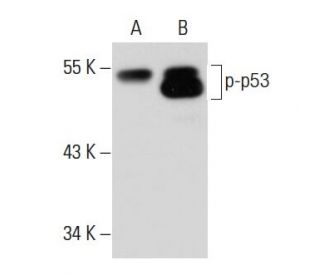

p-p53 Antibody (3H2806): sc-71786
- p-p53 Antibody (3H2806) is a mouse monoclonal IgG1, cited in 5 publications, provided at 100 µg/ml
- raised against a C-terminal phosphopeptide corresponding to amino acids Ser 392 of p53 of human origin
- recommended for detection of Ser 392 phosphorylated p53 of mouse, rat, human, porcine and canine origin by WB, IP, IF, IHC(P) and ELISA; not recommended for detection of dephosphorylated p53
- At present, we have not yet completed the identification of the preferred secondary detection reagent(s) for p-p53 Antibody (3H2806). This work is in progress.

QUICK LINKS
SEE ALSO...
p-p53 Antibody (3H2806) is a mouse monoclonal IgG1 antibody that detects p53 in mouse, rat, human, porcine, and canine samples through various applications including western blotting (WB), immunoprecipitation (IP), immunofluorescence (IF), immunohistochemistry with paraffin-embedded sections (IHCP), and enzyme-linked immunosorbent assay (ELISA). p-p53 monoclonal antibody (3H2806) specifically recognizes the phosphorylated form of p53, a crucial tumor suppressor protein that plays a vital role in cellular stress responses by regulating the expression of genes involved in growth arrest and apoptosis. The phosphorylation of p53 enhances stability and transcriptional activity, allowing p53 to respond to DNA damage and other stress signals. This post-translational modification is essential for activating tumor-suppressive functions, as phosphorylation facilitates the transition from a latent to an active conformation, enabling DNA binding and target gene transcription initiation. Target genes include critical regulators such as p21, which enforces cell cycle arrest by inhibiting cyclin-dependent kinases, and pro-apoptotic factors like BAX and PUMA, which mediate programmed cell death. Through controlling the expression of these downstream effectors, p53 orchestrates a precise cellular response to prevent damaged DNA propagation, highlighting phosphorylation′s importance in maintaining genomic integrity. Since mutations in p53 are frequently associated with various human cancers, detecting and quantifying phosphorylated p53 using anti-p53 antibody (3H2806) is invaluable for research into cancer biology and therapeutic interventions.
Alexa Fluor® is a trademark of Molecular Probes Inc., OR., USA
LI-COR® and Odyssey® are registered trademarks of LI-COR Biosciences
p-p53 Antibody (3H2806) References:
- Effect of Boschniakia rossica on expression of GST-P, p53 and p21(ras)proteins in early stage of chemical hepatocarcinogenesis and its anti-inflammatory activities in rats. | Yin, ZZ., et al. 2000. World J Gastroenterol. 6: 812-818. PMID: 11819701
- WOX1 is essential for tumor necrosis factor-, UV light-, staurosporine-, and p53-mediated cell death, and its tyrosine 33-phosphorylated form binds and stabilizes serine 46-phosphorylated p53. | Chang, NS., et al. 2005. J Biol Chem. 280: 43100-8. PMID: 16219768
- Bcl-2 up-regulation and P-p53 down-regulation account for the low sensitivity of murine L929 fibrosarcoma cells to oridonin-induced apoptosis. | Huang, J., et al. 2005. Biol Pharm Bull. 28: 2068-74. PMID: 16272691
- Dimethylfumarate inhibits MIF-induced proliferation of keratinocytes by inhibiting MSK1 and RSK1 activation and by inducing nuclear p-c-Jun (S63) and p-p53 (S15) expression. | Gesser, B., et al. 2011. Inflamm Res. 60: 643-53. PMID: 21340650
- ATM mediates interdependent activation of p53 and ERK through formation of a ternary complex with p-p53 and p-ERK in response to DNA damage. | Heo, JI., et al. 2012. Mol Biol Rep. 39: 8007-14. PMID: 22576881
- The ATM kinase inhibitor KU-55933 provides neuroprotection against hydrogen peroxide-induced cell damage via a γH2AX/p-p53/caspase-3-independent mechanism: Inhibition of calpain and cathepsin D. | Chwastek, J., et al. 2017. Int J Biochem Cell Biol. 87: 38-53. PMID: 28341201
- Activated mitochondrial apoptosis in hESCs after dissociation involving the PKA/p-p53/Bax signaling pathway. | Zhang, L., et al. 2018. Exp Cell Res. 369: 226-233. PMID: 29802837
- Targeting phosphorylated p53 to elicit tumor-reactive T helper responses against head and neck squamous cell carcinoma. | Ohara, K., et al. 2018. Oncoimmunology. 7: e1466771. PMID: 30510853
- FBXL6 degrades phosphorylated p53 to promote tumor growth. | Li, Y., et al. 2021. Cell Death Differ. 28: 2112-2125. PMID: 33568778
- Nuclear HKII-P-p53 (Ser15) Interaction is a Prognostic Biomarker for Chemoresponsiveness and Glycolytic Regulation in Epithelial Ovarian Cancer. | Han, CY., et al. 2021. Cancers (Basel). 13: PMID: 34298618
Ordering Information
| Product Name | Catalog # | UNIT | Price | Qty | FAVORITES | |
p-p53 Antibody (3H2806) | sc-71786 | 100 µg/ml | $322.00 |