")
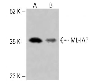
ML-IAP Antibody (E-3): sc-393237
- ML-IAP Antibody (E-3) is a mouse monoclonal IgG1 κ ML-IAP antibody, cited in 1 publications, provided at 200 µg/ml
- specific for an epitope mapping between amino acids 72-97 near the N-terminus of ML-IAP of human origin
- ML-IAP Antibody (E-3) is recommended for detection of ML-IAP of mouse, rat, human origin by WB, IP, IF, IHC(P) and ELISA
- Anti-ML-IAP Antibody (E-3) is available conjugated to agarose for IP; HRP for WB, IHC(P) and ELISA; and to either phycoerythrin or FITC for IF, IHC(P) and FCM
- also available conjugated to Alexa Fluor® 488, Alexa Fluor® 546, Alexa Fluor® 594 or Alexa Fluor® 647 for WB (RGB), IF, IHC(P) and FCM, and for use with RGB fluorescent imaging systems, such as iBright™ FL1000, FluorChem™, Typhoon, Azure and other comparable systems
- also available conjugated to Alexa Fluor® 680 or Alexa Fluor® 790 for WB (NIR), IF and FCM; for use with Near-Infrared (NIR) detection systems, such as LI-COR®Odyssey®, iBright™ FL1000, FluorChem™, Typhoon, Azure and other comparable systems
- m-IgG Fc BP-HRP, m-IgG1 BP-HRP and m-IgGκ BP-HRP are the preferred secondary detection reagents for ML-IAP Antibody (E-3) for WB and IHC(P) applications. These reagents are now offered in bundles with ML-IAP Antibody (E-3) (see ordering information below).

QUICK LINKS
SEE ALSO...
ML-IAP Antibody (E-3) is a mouse monoclonal IgG1 kappa light chain antibody that detects ML-IAP protein of mouse, rat, and human origin by western blotting (WB), immunoprecipitation (IP), immunofluorescence (IF), immunohistochemistry, and enzyme-linked immunosorbent assay (ELISA). ML-IAP (E-3) antibody is available in both non-conjugated and various conjugated forms, including agarose, horseradish peroxidase (HRP), phycoerythrin (PE), fluorescein isothiocyanate (FITC), and multiple Alexa Fluor® conjugates. ML-IAP protein, also known as melanoma inhibitor of apoptosis protein, kidney inhibitor of apoptosis protein (KIAP), livin, or BIRC7, plays a critical role in regulating apoptosis, a vital process for maintaining cellular homeostasis and preventing uncontrolled cell proliferation. ML-IAP contains unique N-terminal baculovirus IAP repeats (BIRs) and a C-terminal RING finger domain, which are essential for function in inhibiting caspases, specifically caspase-9, while binding to caspase-3 and -7 without inhibition. ML-IAP interaction with Smac through BIR domain shows high affinity and specificity, suggesting a complex regulatory mechanism in apoptosis. ML-IAP gene is located on human chromosome 20q13.3, and while debate continues regarding precise localization and role in apoptotic pathways, ML-IAP is increasingly recognized for potential implications in cancer biology and therapeutic resistance.
Alexa Fluor® is a trademark of Molecular Probes Inc., OR., USA
LI-COR® and Odyssey® are registered trademarks of LI-COR Biosciences
ML-IAP Antibody (E-3) References:
- Livin, a novel inhibitor of apoptosis protein family member. | Kasof, GM. and Gomes, BC. 2001. J Biol Chem. 276: 3238-46. PMID: 11024045
- ML-IAP, a novel inhibitor of apoptosis that is preferentially expressed in human melanomas. | Vucic, D., et al. 2000. Curr Biol. 10: 1359-66. PMID: 11084335
- KIAP, a novel member of the inhibitor of apoptosis protein family. | Lin, JH., et al. 2000. Biochem Biophys Res Commun. 279: 820-31. PMID: 11162435
- Two splicing variants of a new inhibitor of apoptosis gene with different biological properties and tissue distribution pattern. | Ashhab, Y., et al. 2001. FEBS Lett. 495: 56-60. PMID: 11322947
- SMAC negatively regulates the anti-apoptotic activity of melanoma inhibitor of apoptosis (ML-IAP). | Vucic, D., et al. 2002. J Biol Chem. 277: 12275-9. PMID: 11801603
- Structure and function analysis of peptide antagonists of melanoma inhibitor of apoptosis (ML-IAP). | Franklin, MC., et al. 2003. Biochemistry. 42: 8223-31. PMID: 12846571
- Identification of an HLA-A3-restricted cytotoxic T lymphocyte (CTL) epitope from ML-IAP. | Andersen, MH., et al. 2004. J Invest Dermatol. 122: 1336-7. PMID: 15140242
- Engineering ML-IAP to produce an extraordinarily potent caspase 9 inhibitor: implications for Smac-dependent anti-apoptotic activity of ML-IAP. | Vucic, D., et al. 2005. Biochem J. 385: 11-20. PMID: 15485396
Ordering Information
| Product Name | Catalog # | UNIT | Price | Qty | FAVORITES | |
ML-IAP Antibody (E-3) | sc-393237 | 200 µg/ml | $322.00 | |||
ML-IAP Antibody (E-3): m-IgG Fc BP-HRP Bundle | sc-530399 | 200 µg Ab; 10 µg BP | $361.00 | |||
ML-IAP Antibody (E-3): m-IgGκ BP-HRP Bundle | sc-523865 | 200 µg Ab, 40 µg BP | $361.00 | |||
ML-IAP Antibody (E-3): m-IgG1 BP-HRP Bundle | sc-544033 | 200 µg Ab; 20 µg BP | $361.00 | |||
ML-IAP Antibody (E-3) AC | sc-393237 AC | 500 µg/ml, 25% agarose | $424.00 | |||
ML-IAP Antibody (E-3) HRP | sc-393237 HRP | 200 µg/ml | $322.00 | |||
ML-IAP Antibody (E-3) FITC | sc-393237 FITC | 200 µg/ml | $336.00 | |||
ML-IAP Antibody (E-3) PE | sc-393237 PE | 200 µg/ml | $349.00 | |||
ML-IAP Antibody (E-3) Alexa Fluor® 488 | sc-393237 AF488 | 200 µg/ml | $364.00 | |||
ML-IAP Antibody (E-3) Alexa Fluor® 546 | sc-393237 AF546 | 200 µg/ml | $364.00 | |||
ML-IAP Antibody (E-3) Alexa Fluor® 594 | sc-393237 AF594 | 200 µg/ml | $364.00 | |||
ML-IAP Antibody (E-3) Alexa Fluor® 647 | sc-393237 AF647 | 200 µg/ml | $364.00 | |||
ML-IAP Antibody (E-3) Alexa Fluor® 680 | sc-393237 AF680 | 200 µg/ml | $364.00 | |||
ML-IAP Antibody (E-3) Alexa Fluor® 790 | sc-393237 AF790 | 200 µg/ml | $364.00 | |||
ML-IAP (E-3) Neutralizing Peptide | sc-393237 P | 100 µg/0.5 ml | $69.00 |