
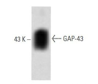

GAP-43 Antibody (31): sc-135915
- GAP-43 Antibody (31) is a mouse monoclonal IgG1, cited in 2 publications, provided at 50 µg/0.5 ml
- raised against amino acids 1-144 of GAP-43 of human origin
- recommended for detection of GAP-43 of mouse, rat and human origin by WB and IP
- See GAP-43 (B-5): sc-17790 for GAP-43 antibody conjugates, including AC, HRP, FITC, PE, Alexa Fluor® 488, 594, 647, 680 and 790.
- At present, we have not yet completed the identification of the preferred secondary detection reagent(s) for GAP-43 Antibody (31). This work is in progress.

QUICK LINKS
SEE ALSO...
GAP-43 Antibody (31) is a mouse monoclonal IgG1 antibody that detects GAP-43 in mouse, rat, and human samples through applications such as western blotting (WB) and immunoprecipitation (IP). GAP-43, also known as growth-associated protein 43, plays a pivotal role in the regenerative response of the nervous system, particularly during neuronal development and axonal regeneration. GAP-43 is predominantly localized in the growth cones of developing neurons, where GAP-43 is essential for guiding axonal growth and facilitating synaptic plasticity. The presence of GAP-43 in these regions underscores GAP-43′s importance in neuronal communication and repair mechanisms. Notably, GAP-43 is regulated by the neuron-specific Elav/Hu family member, HuD, which stabilizes GAP-43 mRNA, leading to increased protein levels crucial for neuronal function. Furthermore, studies have shown that heterozygous GAP-43 knockout mice, which exhibit a 50% reduction in GAP-43 levels, suffer from significant memory impairments, highlighting GAP-43′s critical role in cognitive processes and sensory perception. With an ability to provide insights into neuronal development and regeneration, anti-GAP-43 antibody (31) serves as an invaluable tool for researchers studying neurobiology and related fields.
Alexa Fluor® is a trademark of Molecular Probes Inc., OR., USA
LI-COR® and Odyssey® are registered trademarks of LI-COR Biosciences
GAP-43 Antibody (31) References:
- GAP-43 and neuronal remodeling. | Fishman, MC. and Valenzuela, D. 1991. Prog Brain Res. 89: 89-95. PMID: 1839073
- GAP-43 as a 'calmodulin sponge' and some implications for calcium signalling in axon terminals. | Skene, JH. 1990. Neurosci Res Suppl. 13: S112-25. PMID: 1979675
- Growth-associated Protein-43 (GAP-43) Expression In Ganglionic and Aganglionic Colon. | Grynspan, D., et al. 2012. Pediatr Dev Pathol.. PMID: 22901249
- GAP-43 slows down cell cycle progression via sequences in its 3'UTR. | De Moliner, K., et al. 2015. Arch Biochem Biophys. 571: 66-75. PMID: 25721498
- Retinoic acid protects from experimental cerebral infarction by upregulating GAP-43 expression. | Li, Y., et al. 2017. Braz J Med Biol Res. 50: e5561. PMID: 28380213
- Transient increase in CSF GAP-43 concentration after ischemic stroke. | Sandelius, Å., et al. 2018. BMC Neurol. 18: 202. PMID: 30526557
- GAP-43 is involved in the orientation of cell division by interacting with GΑI during neurogenesis. | Huang, R., et al. 2020. Int J Neurosci. 130: 144-152. PMID: 31554446
- CSF GAP-43 as a biomarker of synaptic dysfunction is associated with tau pathology in Alzheimer's disease. | Qiang, Q., et al. 2022. Sci Rep. 12: 17392. PMID: 36253408
- P53-induced GAP-43 Upregulation in Primary Cortical Neurons of Rats. | Li, T., et al. 2024. Protein Pept Lett. 31: 229-235. PMID: 38288820
- GAP-43, aFGF, CCK and alpha- and beta-CGRP in rat spinal motoneurons subjected to axotomy and/or dorsal root severance. | Piehl, F., et al. 1993. Eur J Neurosci. 5: 1321-33. PMID: 7506107
Ordering Information
| Product Name | Catalog # | UNIT | Price | Qty | FAVORITES | |
GAP-43 Antibody (31) | sc-135915 | 50 µg/0.5 ml | $322.00 |