")
AK2 Antibody (F-2): sc-374095
- AK2 Antibody (F-2) is a mouse monoclonal IgG1 κ AK2 antibody, cited in 5 publications, provided at 200 µg/ml
- raised against amino acids 31-95 mapping near the N-terminus of AK2 of human origin
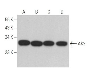
- AK2 Antibody (F-2) is recommended for detection of AK2 of mouse, rat and human origin by WB, IP, IF, IHC(P) and ELISA
- Anti-AK2 Antibody (F-2) is available conjugated to agarose for IP; HRP for WB, IHC(P) and ELISA; and to either phycoerythrin or FITC for IF, IHC(P) and FCM
- also available conjugated to Alexa Fluor® 488, Alexa Fluor® 546, Alexa Fluor® 594 or Alexa Fluor® 647 for WB (RGB), IF, IHC(P) and FCM, and for use with RGB fluorescent imaging systems, such as iBright™ FL1000, FluorChem™, Typhoon, Azure and other comparable systems
- also available conjugated to Alexa Fluor® 680 or Alexa Fluor® 790 for WB (NIR), IF and FCM; for use with Near-Infrared (NIR) detection systems, such as LI-COR®Odyssey®, iBright™ FL1000, FluorChem™, Typhoon, Azure and other comparable systems
- m-IgG Fc BP-HRP, m-IgG1 BP-HRP and m-IgGκ BP-HRP are the preferred secondary detection reagents for AK2 Antibody (F-2) for WB and IHC(P) applications. These reagents are now offered in bundles with AK2 Antibody (F-2) (see ordering information below).

AK2 Antibody (F-2) is a mouse monoclonal IgG1 kappa light chain antibody that detects AK2 protein of mouse, rat, and human origin by western blotting (WB), immunoprecipitation (IP), immunofluorescence (IF), immunohistochemistry, and enzyme-linked immunosorbent assay (ELISA). AK2 Antibody (F-2) is available in both non-conjugated and various conjugated forms, including agarose, horseradish peroxidase (HRP), phycoerythrin (PE), fluorescein isothiocyanate (FITC), and multiple Alexa Fluor® conjugates. AK2 plays a crucial role in cellular energy metabolism through involvement in adenine nucleotide phosphorylation, essential for energy transfer within cells. AK2 (F-2) antibody targets proteins localized in mitochondrial intermembrane space, where AK2 participates in ATP level regulation, influencing metabolic pathways and cellular signaling. This specific localization enables rapid response to changes in cellular energy demands, especially during stress conditions such as apoptosis, where AK2 translocates to the cytosol alongside cytochrome c. AK2′s movement from mitochondria to cytosol during early apoptotic events suggests a role in cell death pathways and highlights mitochondrial function in maintaining cellular homeostasis.
Alexa Fluor® is a trademark of Molecular Probes Inc., OR., USA
LI-COR® and Odyssey® are registered trademarks of LI-COR Biosciences
AK2 Antibody (F-2) References:
- Release of adenylate kinase 2 from the mitochondrial intermembrane space during apoptosis. | Köhler, C., et al. 1999. FEBS Lett. 447: 10-2. PMID: 10218571
- Adenylate kinase phosphotransfer communicates cellular energetic signals to ATP-sensitive potassium channels. | Carrasco, AJ., et al. 2001. Proc Natl Acad Sci U S A. 98: 7623-8. PMID: 11390963
- Adenylate kinase 2, a mitochondrial enzyme. | Bruns, GA. and Regina, VM. 1977. Biochem Genet. 15: 477-86. PMID: 195572
- Cloning and characterization of cDNA for human adenylate kinase 2A. | Lee, Y., et al. 1996. Biochem Mol Biol Int. 39: 833-42. PMID: 8843353
- cDNA cloning and tissue-specific expression of the gene encoding human adenylate kinase isozyme 2. | Noma, T., et al. 1998. Biochim Biophys Acta. 1395: 34-9. PMID: 9434148
- Cloning and expression of human adenylate kinase 2 isozymes: differential expression of adenylate kinase 1 and 2 in human muscle tissues. | Lee, Y., et al. 1998. J Biochem. 123: 47-54. PMID: 9504408
- Adenylate kinase: kinetic behavior in intact cells indicates it is integral to multiple cellular processes. | Dzeja, PP., et al. 1998. Mol Cell Biochem. 184: 169-82. PMID: 9746320
Ordering Information
| Product Name | Catalog # | UNIT | Price | Qty | FAVORITES | |
AK2 Antibody (F-2) | sc-374095 | 200 µg/ml | $322.00 | |||
AK2 Antibody (F-2): m-IgG Fc BP-HRP Bundle | sc-529658 | 200 µg Ab; 10 µg BP | $361.00 | |||
AK2 Antibody (F-2): m-IgGκ BP-HRP Bundle | sc-522619 | 200 µg Ab, 40 µg BP | $361.00 | |||
AK2 Antibody (F-2): m-IgG1 BP-HRP Bundle | sc-543626 | 200 µg Ab; 20 µg BP | $361.00 | |||
AK2 Antibody (F-2) AC | sc-374095 AC | 500 µg/ml, 25% agarose | $424.00 | |||
AK2 Antibody (F-2) HRP | sc-374095 HRP | 200 µg/ml | $322.00 | |||
AK2 Antibody (F-2) FITC | sc-374095 FITC | 200 µg/ml | $336.00 | |||
AK2 Antibody (F-2) PE | sc-374095 PE | 200 µg/ml | $349.00 | |||
AK2 Antibody (F-2) Alexa Fluor® 488 | sc-374095 AF488 | 200 µg/ml | $364.00 | |||
AK2 Antibody (F-2) Alexa Fluor® 546 | sc-374095 AF546 | 200 µg/ml | $364.00 | |||
AK2 Antibody (F-2) Alexa Fluor® 594 | sc-374095 AF594 | 200 µg/ml | $364.00 | |||
AK2 Antibody (F-2) Alexa Fluor® 647 | sc-374095 AF647 | 200 µg/ml | $364.00 | |||
AK2 Antibody (F-2) Alexa Fluor® 680 | sc-374095 AF680 | 200 µg/ml | $364.00 | |||
AK2 Antibody (F-2) Alexa Fluor® 790 | sc-374095 AF790 | 200 µg/ml | $364.00 |