")
XPA Antikörper (B-1): sc-28353
- XPA Antikörper B-1 ist ein Maus monoklonales IgG1 κ XPA Antikörper, verwendet in 26 wissenschaftlichen Veröffentlichungen, in einer Menge von 200 µg/ml
- gezogen gegen Aminosäuren 1-273, die das Volllänge- XPA aus der Spezies human darstellen
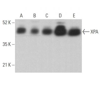
- XPA Antikörper (B-1) ist empfohlen für die Detektion von XPA aus der Spezies mouse, rat und human per WB, IP, IF, IHC(P) und ELISA
- Anti-XPA Antikörper (B-1) ist erhältlich als Konjugat mit Agarose für IP; HRP für WB, IHC(P) und ELISA; und entweder mit Phycoerythrin oder FITC für IF, IHC(P) und FCM
- auch erhältlich als Konjugat mit Alexa Fluor® 488, Alexa Fluor® 546, Alexa Fluor® 594 oder Alexa Fluor® 647 für IF, IHC(P) und FCM
- auch erhältlich als Konjugat mit Alexa Fluor® 680 oder Alexa Fluor® 790 für WB (NIR), IF und FCM
- m-IgG Fc BP-HRP, 1 BP-HRP">m-IgG1 BP-HRP und m-IgGκ BP-HRP sind die bevorzugten sekundären Nachweisreagenzien für XPA Antikörper (B-1) für WB- und IHC(P)-Anwendungen. Diese Reagenzien werden jetzt in Bündeln mit XPA Antikörper (B-1) angeboten(siehe Bestellinformationen unten).

Direktverknüpfungen
Siehe auch...
Der XPA-Antikörper (B-1) ist ein monoklonaler IgG1-Antikörper der leichten Kette Kappa aus Mäusen, der das XPA-Protein von Mäusen, Ratten und Menschen durch Western Blot (WB), Immunopräzipitation (IP), Immunfluoreszenz (IF), Immunhistochemie und Enzyme-Linked Immunosorbent Assay (ELISA) nachweist. Der anti-XPA-Antikörper (B-1) ist sowohl in nicht konjugierter als auch in verschiedenen konjugierten Formen erhältlich, darunter Agarose, Meerrettichperoxidase (HRP), Phycoerythrin (PE), Fluoresceinisothiocyanat (FITC) und mehrere Alexa Fluor®-Konjugate. Das XPA-Protein spielt eine entscheidende Rolle im Nukleotid-Exzisionsreparatur-Signalweg (NER), der für die Reparatur von DNA-Schäden durch ultraviolettes (UV) Licht unerlässlich ist. Diese Funktion ist besonders wichtig, da Defizite in der NER zu Xeroderma pigmentosum führen können, einer genetischen Störung, die das Hautkrebsrisiko aufgrund der Anhäufung nicht reparierter DNA-Läsionen erheblich erhöht. XPA ist für die Koordination der Zusammensetzung des Präinkisionskomplexes verantwortlich, der für die Erkennung und Entfernung beschädigter DNA erforderlich ist, und XPA weist eine einzigartige Fähigkeit auf, sich sowohl an einzelsträngige als auch an doppelsträngige DNA zu binden, wodurch die Effizienz bei der Schadenserkennung erhöht wird. Darüber hinaus unterstreicht die Fähigkeit von XPA, in Abwesenheit von DNA Homodimere zu bilden und bei Bindung an DNA in eine wirksamere dimere Form überzugehen, die Regulationsmechanismen, die die Aktivität als Reaktion auf DNA-Schäden steuern. Die Bedeutung von XPA für die Aufrechterhaltung der genomischen Integrität unterstreicht das Potenzial als Ziel für therapeutische Interventionen bei Erkrankungen, die mit gestörten DNA-Reparaturmechanismen in Verbindung stehen.
Alexa Fluor® ist ein Markenzeichen von Molecular Probes Inc., OR., USA
LI-COR® und Odyssey® sind Markenzeichen von LI-COR Biosciences
XPA Antikörper (B-1) Literaturhinweise:
- Die relative Expression der mutierten XPB-Gene führt zu den zellulären Phänotypen Xeroderma pigmentosum/Cockayne-Syndrom oder Trichothiodystrophie. | Riou, L., et al. 1999. Hum Mol Genet. 8: 1125-33. PMID: 10332046
- Photobiologische und photoimmunologische Eigenschaften von Mäusen mit XPA-Genmangel. | Horio, T., et al. 2001. J Investig Dermatol Symp Proc. 6: 58-63. PMID: 11764287
- Das Xeroderma-Pigmentosum-Komplementationsgruppe-A-Protein (XPA) moduliert die RPA-DNA-Interaktionen durch erhöhte Komplexstabilität und Hemmung der Strangtrennungsaktivität. | Patrick, SM. and Turchi, JJ. 2002. J Biol Chem. 277: 16096-101. PMID: 11859086
- Die kooperative Interaktion von menschlichem XPA stabilisiert und verstärkt die spezifische Bindung von XPA an DNA-Schäden. | Liu, Y., et al. 2005. Biochemistry. 44: 7361-8. PMID: 15882075
- Gegensätzliche Auswirkungen des UV-Läsionsreparaturproteins XPA und der UV-Bypass-Polymerase eta auf die ATR-Checkpoint-Signalgebung. | Bomgarden, RD., et al. 2006. EMBO J. 25: 2605-14. PMID: 16675950
- ATR-abhängiger Kontrollpunkt moduliert den XPA-Kernimport als Reaktion auf UV-Bestrahlung. | Wu, X., et al. 2007. Oncogene. 26: 757-64. PMID: 16862173
- XPA-Mangel beeinträchtigt die Funktion des Ubiquitin-Proteasom-Systems. | de Sousa Leal, AM., et al. 2020. DNA Repair (Amst). 94: 102937. PMID: 32693352
- Erkennung von Läsionen durch XPC, TFIIH und XPA bei der DNA-Exzisionsreparatur. | Kim, J., et al. 2023. Nature. 617: 170-175. PMID: 37076618
- Artenübergreifende Untersuchung der Notwendigkeit von XPA für die Nukleotid-Exzisionsreparatur. | Kose, C., et al. 2024. Nucleic Acids Res. 52: 677-689. PMID: 37994737
- Hohes Auftreten von Hauttumoren, die durch Ultraviolett-B-Strahlen oder chemische Karzinogene ausgelöst werden, bei Mäusen, denen das Gen für Xeroderma pigmentosum Gruppe A fehlt. | Nakane, H., et al. 1995. Nature. 377: 165-8. PMID: 7675085
- Identifizierung einer Bindungsdomäne für beschädigte DNA des XPA-Proteins. | Kuraoka, I., et al. 1996. Mutat Res. 362: 87-95. PMID: 8538652
- Trennung der Proteinfaktoren, die die Defekte in den sieben Komplementationsgruppen der Xeroderma-Pigmentosum-Zellen korrigieren. | Tateishi, S., et al. 1995. J Biochem. 118: 819-24. PMID: 8576098
Bestellinformation
| Produkt | Katalog # | EINHEIT | Preis | ANZAHL | Favoriten | |
XPA Antikörper (B-1) | sc-28353 | 200 µg/ml | $322.00 | |||
XPA (B-1): m-IgG Fc BP-HRP Bundle | sc-528432 | 200 µg Ab; 10 µg BP | $361.00 | |||
XPA (B-1): m-IgGκ BP-HRP Bundle | sc-520807 | 200 µg Ab, 40 µg BP | $361.00 | |||
XPA (B-1): m-IgG1 BP-HRP Bundle | sc-542931 | 200 µg Ab; 20 µg BP | $361.00 | |||
XPA Antikörper (B-1) AC | sc-28353 AC | 500 µg/ml, 25% agarose | $424.00 | |||
XPA Antikörper (B-1) HRP | sc-28353 HRP | 200 µg/ml | $322.00 | |||
XPA Antikörper (B-1) FITC | sc-28353 FITC | 200 µg/ml | $336.00 | |||
XPA Antikörper (B-1) PE | sc-28353 PE | 200 µg/ml | $349.00 | |||
XPA Antikörper (B-1) Alexa Fluor® 488 | sc-28353 AF488 | 200 µg/ml | $364.00 | |||
XPA Antikörper (B-1) Alexa Fluor® 546 | sc-28353 AF546 | 200 µg/ml | $364.00 | |||
XPA Antikörper (B-1) Alexa Fluor® 594 | sc-28353 AF594 | 200 µg/ml | $364.00 | |||
XPA Antikörper (B-1) Alexa Fluor® 647 | sc-28353 AF647 | 200 µg/ml | $364.00 | |||
XPA Antikörper (B-1) Alexa Fluor® 680 | sc-28353 AF680 | 200 µg/ml | $364.00 | |||
XPA Antikörper (B-1) Alexa Fluor® 790 | sc-28353 AF790 | 200 µg/ml | $364.00 |