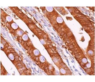
Date published: 2025-9-7

00800 4573 8000

SCBT Portrait Logo
Seach Input

TBK1 Antikörper (A-6): sc-398366

3.6(9)
Produkt bewertenBitte stellen Sie eine Frage

Datenblätter
  • TBK1 Antikörper A-6 ist ein Maus monoklonales IgG1 κ TBK1 Antikörper, verwendet in 12 wissenschaftlichen Veröffentlichungen, in einer Menge von 200 µg/ml
  • gezogen gegen die Aminosäuresequenz 355-729 lokalisiert am C-terminus von TBK1 aus der Spezies mouse
  • TBK1 Antikörper (A-6) ist empfohlen für die Detektion von TBK1 aus der Spezies mouse, rat und human per WB, IP, IF, IHC(P) und ELISA
  • Anti-TBK1 Antikörper (A-6) ist erhältlich als Konjugat mit Agarose für IP; HRP für WB, IHC(P) und ELISA; und entweder mit Phycoerythrin oder FITC für IF, IHC(P) und FCM
  • auch erhältlich als Konjugat mit Alexa Fluor® 488, Alexa Fluor® 546, Alexa Fluor® 594 oder Alexa Fluor® 647 für IF, IHC(P) und FCM
  • auch erhältlich als Konjugat mit Alexa Fluor® 680 oder Alexa Fluor® 790 für WB (NIR), IF und FCM
  • Bitte kontaktieren Sie uns um ein 10 µg GRATIS Sample vom TBK1 (A-6): sc-398366 zu erhalten.
  • m-IgG Fc BP-HRP, m-IgG1 BP-HRP und m-IgGκ BP-HRP sind die empfohlenen Detektionsreagenzien für TBK1 Antikörper (A-6) für WB and IHC(P) Experimente. Diese Reagenzien werden nun auch als Bundle angeboten, zusammen mit TBK1 Antikörper (A-6) (Bestellinformation unten stehend).

Direktverknüpfungen

Siehe auch...

Der TBK1-Antikörper (A-6) ist ein monoklonaler Maus IgG1 κ TBK1-Antikörper (auch als NAK-Antikörper, T2K-Antikörper, Encephalopathie, akut, infektionsbedingt, herpes-spezifisch, 8 (IIAE8) Antikörper oder Frontotemporale Demenz und/oder Amyotrophe Lateralsklerose 4 (FTDALS4) Antikörper bezeichnet), der das TBK1-Protein von Maus-, Ratte- und menschlicher Herkunft mittels WB, IP, IF, IHC(P) und ELISA detektiert. Der TBK1-Antikörper (A-6) ist sowohl in nicht konjugierter Form als auch in mehreren konjugierten Formen des TBK1-Antikörpers erhältlich, darunter Agarose, HRP, PE, FITC und mehrere Alexa Fluor®-Konjugate. Der Transkriptionsfaktor NFkB wird im Zytoplasma in einer inaktiven Form durch das inhibitory Protein IkB gehalten. Die Aktivierung von NFkB erfordert, dass IkB an bestimmten Serinresten phosphoryliert wird, was zu einer gezielten Degradation von IkB führt. IkB-Kinase α (IKKα), früher als CHUK bezeichnet, interagiert mit IkB-a und phosphoryliert spezifisch IkB-α an den Stellen, die seine Degradation auslösen, Serin 32 und 36. Das funktionelle IKK-Komplex enthält drei Untereinheiten, IKKα, IKKβ und IKKγ (auch als NEMO bezeichnet), und jede scheint einen essentiellen Beitrag zur Phosphorylierung von IkB zu leisten. Die TANK-Bindungskinase (TBK1), auch als T2K bezeichnet, ist eine neuartige IKK-verwandte Kinase, die in murinen und menschlichen Geweben identifiziert wurde. TBK1 wurde gezeigt, dass es sich mit TRAF2 und TANK in der NFkB-Aktivierungspathway komplexiert. TBK1 weist eine Homologie mit IKKα und IKKβ im Amino-Terminalteil auf, der die Kinase-Domäne einschließt.

Ausschließlich für Forschungszwecke. Nicht für diagnostische oder therapeutische Zwecke bestimmt.

Alexa Fluor® ist ein Markenzeichen von Molecular Probes Inc., OR., USA

LI-COR® und Odyssey® sind Markenzeichen von LI-COR Biosciences

TBK1 Antikörper (A-6) Literaturhinweise:

  1. STING spezifiziert die IRF3-Phosphorylierung durch TBK1 im zytosolischen DNA-Signalweg.  |  Tanaka, Y. and Chen, ZJ. 2012. Sci Signal. 5: ra20. PMID: 22394562
  2. TRIM23 vermittelt die virusinduzierte Autophagie über die Aktivierung von TBK1.  |  Sparrer, KMJ., et al. 2017. Nat Microbiol. 2: 1543-1557. PMID: 28871090
  3. Raum-zeitliche Kontrolle der ULK1-Aktivierung durch NDP52 und TBK1 während der selektiven Autophagie.  |  Vargas, JNS., et al. 2019. Mol Cell. 74: 347-362.e6. PMID: 30853401
  4. Die Rekrutierung von TBK1 zu STING aktiviert sowohl IRF3 als auch NF-κB, die die Immunabwehr gegen Tumore und Virusinfektionen vermitteln.  |  Yum, S., et al. 2021. Proc Natl Acad Sci U S A. 118: PMID: 33785602
  5. Tilirosid zielt auf TBK1, um Ferroptose zu induzieren und hepatozelluläre Karzinome für Sorafenib zu sensibilisieren.  |  Yang, C., et al. 2023. Phytomedicine. 111: 154668. PMID: 36657316
  6. VANGL2 hemmt die antivirale IFN-I-Signalisierung, indem es TBK1 für den autophagischen Abbau anvisiert.  |  Hu, Z., et al. 2023. Sci Adv. 9: eadg2339. PMID: 37352355
  7. ATG4B hemmt die antivirale Immunität durch GABARAP-gesteuerten autophagischen Abbau von TBK1.  |  Xie, W., et al. 2023. Autophagy. 19: 2853-2868. PMID: 37434364
  8. Regulierung des Transferrinrezeptor-Transports durch Optineurin und seine krankheitsassoziierten Mutanten.  |  Yamano, K., et al. 2024. EMBO J. 43: 754-779. PMID: 38287189
  9. Eine TBK1-Variante verursacht bei Mäusen Autophagolysosomal- und Motoneuronpathologie ohne Neuroinflammation.  |  Brenner, D., et al. 2024. J Exp Med. 221: PMID: 38517332
  10. CDK4/6 verändert die TBK1-Phosphorylierung, um den STING-Signalweg bei Prostatakrebs zu hemmen.  |  Li, W., et al. 2024. Cancer Res. 84: 2588-2606. PMID: 38861362

Bestellinformation

ProduktKatalog #EINHEITPreisANZAHLFavoriten

TBK1 Antikörper (A-6)

sc-398366
200 µg/ml
$316.00

TBK1 (A-6): m-IgG Fc BP-HRP Bundle

sc-530775
200 µg Ab; 10 µg BP
$354.00

TBK1 (A-6): m-IgGκ BP-HRP Bundle

sc-524312
200 µg Ab, 40 µg BP
$354.00

TBK1 (A-6): m-IgG1 BP-HRP Bundle

sc-544247
200 µg Ab; 20 µg BP
$354.00

TBK1 Antikörper (A-6) AC

sc-398366 AC
500 µg/ml, 25% agarose
$416.00

TBK1 Antikörper (A-6) HRP

sc-398366 HRP
200 µg/ml
$316.00

TBK1 Antikörper (A-6) FITC

sc-398366 FITC
200 µg/ml
$330.00

TBK1 Antikörper (A-6) PE

sc-398366 PE
200 µg/ml
$343.00

TBK1 Antikörper (A-6) Alexa Fluor® 488

sc-398366 AF488
200 µg/ml
$357.00

TBK1 Antikörper (A-6) Alexa Fluor® 546

sc-398366 AF546
200 µg/ml
$357.00

TBK1 Antikörper (A-6) Alexa Fluor® 594

sc-398366 AF594
200 µg/ml
$357.00

TBK1 Antikörper (A-6) Alexa Fluor® 647

sc-398366 AF647
200 µg/ml
$357.00

TBK1 Antikörper (A-6) Alexa Fluor® 680

sc-398366 AF680
200 µg/ml
$357.00

TBK1 Antikörper (A-6) Alexa Fluor® 790

sc-398366 AF790
200 µg/ml
$357.00

How can TBK1 (A-6): sc-398366 monoclonal antibody be used for double or triple staining, if the other primary antibodies are also raised in mouse?

Gefragt von: jenni
Thank you for your question In this case, we recommend using a directly conjugated mouse monoclonal primary antibody. Please contact our special orders department for pricing and availability of sc-398366 conjugates.
Beantwortet von: Santa Cruz TS
Veröffentlichungsdatum: 2017-01-30
  • y_2025, m_8, d_29, h_8CST
  • bvseo_bulk, prod_bvqa, vn_bulk_3.0.42
  • cp_1, bvpage1
  • co_hasquestionsanswers, tq_1
  • loc_de_DE, sid_398366, prod, sort_[SortEntry(order=LAST_APPROVED_ANSWER_SUBMISSION_TIME, direction=DESCENDING)]
  • clientName_scbt
  • bvseo_sdk, java_sdk, bvseo-4.0.0
  • CLOUD, getContent, 102ms
  • QUESTIONS, PRODUCT
Rated 4 von 5 von aus GoodThis antibody recognizes a protein of the right molecular weigh, though the signal is weak in western blot of HL60, Jurkat and K562 cells
Veröffentlichungsdatum: 2021-05-31
Rated 3 von 5 von aus Low signal in WB analysis for murine cell lineI use 1:500 diluition in WB and a low signal was detected in J774 cell line
Veröffentlichungsdatum: 2020-11-18
Rated 4 von 5 von aus Good antibodyThis antibody worked very well with fixed frozen mouse tissue sections for IF
Veröffentlichungsdatum: 2018-10-03
Rated 1 von 5 von aus Not specificWe bought 2 TBK1 antibodies recently, sc-398366 and sc-52957. The first one works for blotting in mouse cells but NOT in human cells where it sees an irrelevant band of higher MW which does not go away with siRNA. The second one works in both mouse and human cells and the band that it sees is correct and goes away with siRNA. So the band shown for HeLa cells is rightly not the right size, and not a smiling gel. It will be best and more believable if Santa Cruz staff did not post reviews unless sure about what they are saying.
Veröffentlichungsdatum: 2017-10-13
Rated 5 von 5 von aus Great!Works well with single band at the expected size! Used in western blot in 1:1000 dilution on a lysate from HepG2 cells.
Veröffentlichungsdatum: 2017-09-13
Rated 1 von 5 von aus It is not working in liver cancer cell linesI got free samples recently and did western blot in liver cancer cell lines HepG2 and Snu449 after knockdown. The antibody picked up one band with bigger size than prediction. Also, there is no change for the band density after knockdown.
Veröffentlichungsdatum: 2017-04-28
Rated 5 von 5 von aus Well characterized TBK1 ab for WB & IHCBeautiful cytoplasmic Immunoperoxidase staining on formalin fixed, paraffin-embedded human duodenum tissue and human adrenal gland tissue.
Veröffentlichungsdatum: 2017-01-14
Rated 4 von 5 von aus Potent and very specific antibodyThis monoclonal antibody was independently tested in-house by Kinexus Bioinformatics Corporation and validated for use for Western blotting. There was medium detection of the ~80 kDa target protein in mouse tissues (brain/testes/thymus/muscle mix). No major cross-reactivities were observed at 2 µg/ml antibody concentration with off-target proteins in the mouse tissues tested.
Veröffentlichungsdatum: 2016-11-10
  • y_2025, m_8, d_29, h_8
  • bvseo_bulk, prod_bvrr, vn_bulk_3.0.42
  • cp_1, bvpage1
  • co_hasreviews, tv_0, tr_9
  • loc_de_DE, sid_398366, prod, sort_[SortEntry(order=SUBMISSION_TIME, direction=DESCENDING)]
  • clientName_scbt
  • bvseo_sdk, java_sdk, bvseo-4.0.0
  • CLOUD, getReviews, 14ms
  • REVIEWS, PRODUCT
TBK1 Antikörper (A-6) wurde bewertet mit 3.6 von 5 von 9.
  • y_2025, m_8, d_29, h_8
  • bvseo_bulk, prod_bvrr, vn_bulk_3.0.42
  • cp_1, bvpage1
  • co_hasreviews, tv_0, tr_9
  • loc_de_DE, sid_398366, prod, sort_[SortEntry(order=SUBMISSION_TIME, direction=DESCENDING)]
  • clientName_scbt
  • bvseo_sdk, java_sdk, bvseo-4.0.0
  • CLOUD, getAggregateRating, 105ms
  • REVIEWS, PRODUCT