")
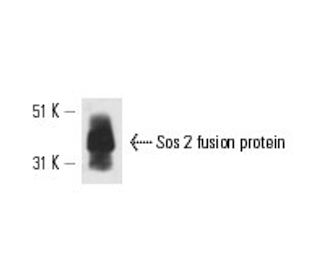
: sc-55478. Análisis Western blot de la proteína de fusión Sos 2 recombinante humana.")
Sos 2 Anticuerpo (A-8): sc-55478
- Sos 2 Anticuerpo (A-8) es un monoclonal de ratón IgG2a (kappa light chain) proporcionado como 200 µg/ml
- Producido contra los amino ácidos 1091-1170 cerca de el C-terminus de Sos 2 de origen human
- recomendado para detectar Sos 2 de human origen, mediante WB, IP, IF y ELISA
- m-IgG Fc BP-HRP es el reactivo de detección secundario preferido para Sos 2 Anticuerpo (A-8) para aplicaciones WB. Este reactivo se ofrece ahora en un paquete con Sos 2 Anticuerpo (A-8)(ver información de pedido más abajo).

ENLACES RÁPIDOS
La super familia de proteínas de unión a GTP, de las cuales las proteínas Ras son prototipos, ha sido implicada en una amplia gama de actividades biológicas. Estudios han identificado una familia de factores liberadores de nucleótidos de guanina (GRFs) que activan Ras en células de mamíferos y una proteína "adaptadora" (Sem 5/GRB2) que parece mediar la interacción de GRFs con moléculas receptoras activadas. Ras-GRF p140 promueve el intercambio de nucleótidos en Ras p21s pero no en otros miembros de la super familia de genes Ras. Además, se han descrito tres homólogos mamíferos del Ras-GRF de Drosophila, hijo de sieteless (Sos). Estos incluyen dos de ratón, mSos 1 y mSos 2, y uno de humano, hSos. Se ha informado que Vav p95 funciona como un GRF en la activación de Ras por el receptor de células T y se ha informado que tiene un dominio similar al de Dbl p115, que es un GRF específico para Cdc42Hs. Posterior a la activación, Ras parece interactuar con Raf, activando así la vía de fosforilación de la MAP quinasa.
Alexa Fluor® es una marca registrada de Molecular Probes Inc., OR., USA
REIVEW LI-COR® y Odyssey® son marcas registradas de LI-COR Biosciences.
Sos 2 Anticuerpo (A-8) Referencias:
- Estudio de asociación de genes candidatos de los genes de señalización de la insulina y la enfermedad de Alzheimer: pruebas de SOS2, PCK1 y PPARgamma como loci de susceptibilidad. | Hamilton, G., et al. 2007. Am J Med Genet B Neuropsychiatr Genet. 144B: 508-16. PMID: 17440948
- La activación de la señalización Ras por Francisella tularensis intracelular mediante el reclutamiento de PKCα y βI al complejo SOS2/GrB2 es esencial para la proliferación bacteriana en el citosol. | Al-Khodor, S. and Abu Kwaik, Y. 2010. Cell Microbiol. 12: 1604-21. PMID: 20618341
- Variantes raras en SOS2 y LZTR1 están asociadas con el síndrome de Noonan. | Yamamoto, GL., et al. 2015. J Med Genet. 52: 413-21. PMID: 25795793
- Los loci SOS2 y ACP1 identificados mediante el análisis de chips de exoma a gran escala regulan el desarrollo y la función renal. | Li, M., et al. 2017. J Am Soc Nephrol. 28: 981-994. PMID: 27920155
- Los FMAM SOS en la salud y la enfermedad. | Baltanás, FC., et al. 2020. Biochim Biophys Acta Rev Cancer. 1874: 188445. PMID: 33035641
- Primer caso prenatal de síndrome de Noonan con mutación SOS2: Implicaciones del diagnóstico precoz para el consejo genético. | Gentile, M., et al. 2021. Am J Med Genet A. 185: 1897-1902. PMID: 33750022
- SOS2 entra en escena: Funcionalidades diferenciales en fisiología y patología. | Baltanás, FC., et al. 2021. Int J Mol Sci. 22: PMID: 34205562
- El sulfaroteno, un retinoide sintético, supera la resistencia al sorafenib del carcinoma hepatocelular a través de la supresión de la vía SOS2-RAS. | Qi, F., et al. 2021. J Exp Clin Cancer Res. 40: 280. PMID: 34479623
- Anomalías linfáticas a lo largo de la vida en pacientes con síndrome de Noonan: Estudio de cohortes retrospectivo. | Swarts, JW., et al. 2022. Am J Med Genet A. 188: 3242-3261. PMID: 35979676
- Localización cromosómica de dos genes que codifican factores de intercambio ras humanos: SOS1 se localiza en la región 2p22-->p16 y SOS2 en la región 14q21-->q22 del genoma humano. | Chardin, P. and Mattei, MG. 1994. Cytogenet Cell Genet. 66: 68-9. PMID: 8275713
Información sobre pedidos
| Nombre del producto | Número de catálogo | UNIDAD | Precio | CANTIDAD | Favoritos | |
Sos 2 Anticuerpo (A-8) | sc-55478 | 200 µg/ml | $322.00 | |||
Paquete de Sos 2 (A-8): m-IgG Fc BP-HRP | sc-539020 | 200 µg Ab; 10 µg BP | $361.00 |