Date published: 2025-10-27

00800 4573 8000

SCBT Portrait Logo
Seach Input

RUNX1 Antikörper (DW71): sc-101146

5.0(10)
Produkt bewertenBitte stellen Sie eine Frage

Datenblätter
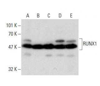
  • RUNX1 Antikörper (DW71) ist ein Maus monoklonales IgG2b κ, verwendet in 15 wissenschaftlichen Veröffentlichungen, in einer Menge von 100 µg/ml
  • gezogen gegen rekombinantes RUNX1 aus der Spezies human
  • Empfohlen für die Detektion von a broad range of RUNX1 isoforms aus der Spezies mouse, rat und human per WB, IP, IF, IHC(P) und ELISA
  • Als Direktkonjugat zur Detektion von RUNX1 wird RUNX1 (A-2): sc-365644 angeboten; Primärantikörper konjugiert mit AC, HRP, FITC, PE, Alexa Fluor® 488, 594, 647, 680 und 790.
  • Aktuell testen wir noch unsere Sekundärantikörper um das beste Bindeprotein für diesen Primärantikörper RUNX1 (DW71) zu finden. Kontaktieren Sie uns bitte, wenn Sie Fragen hierzu haben sollten.

Direktverknüpfungen

Siehe auch...

Der RUNX1-Antikörper (DW71) ist ein monoklonaler Maus IgG2b κ RUNX1-Antikörper, der durch WB, IP, IF, IHC (P) und ELISA eine breite Palette von RUNX1-Isoformen aus Maus-, Ratte- und menschlicher Herkunft detektiert. Der RUNX1-Antikörper (DW71) ist als nicht konjugierte Form des Anti-RUNX1-Antikörpers erhältlich. Die Säugetier-Runt-verwandte Transkriptionsfaktorfamilie (RUNX) besteht aus drei Mitgliedern, RUNX1 (auch als AML-1, PEBP2αB, CBFA2 bezeichnet), RUNX2 (auch als AML-3, PEBP2αA, CBFA1, Osf2 bezeichnet) und RUNX3 (auch als AML-2, PEBPαC, CBFA3 bezeichnet). RUNX-Familienmitglieder sind DNA-bindende Proteine, die die Expression von Genen regulieren, die an der Zelldifferenzierung und Zellzyklusprogression beteiligt sind. RUNX1 ist an der Hämatopoese beteiligt und wird häufig durch chromosomale Translokationen in menschlichen Leukämien als Zielwert anvisiert, bei denen die DNA-bindende Domäne von RUNX1 mit anderen Transkriptionsfaktoren und Corepressormolekülen fusioniert wird. Neben seiner Rolle in der Leukämogenese ist RUNX1 auch an der Differenzierung sensorischer Neuronen beteiligt. Insbesondere fördert RUNX1 die axonale Wachstumsrate, wird selektiv in neuralen Kammzellen abgeleiteten TrkA+-sensorischen Neuronen exprimiert und vermittelt die TrkA-Transaktivierung in migratorischen neuralen Kammzellen. Alternatives Splicing führt zu mehreren Isoformen von RUNX1.

Ausschließlich für Forschungszwecke. Nicht für diagnostische oder therapeutische Zwecke bestimmt.

Bestellinformation

ProduktKatalog #EINHEITPreisANZAHLFavoriten

RUNX1 Antikörper (DW71)

sc-101146
100 µg/ml
$333.00

Hello, is this antibody recognize the N terminal or C terminal of RUNX1?

Gefragt von: YIZHEN
Thank you for your question. This RUNX1 antibody was raised against recombinant protein mapping within amino acids 210-310 of RUNX1 of human origin. The exact epitope has not been mapped.
Beantwortet von: Tech Service
Veröffentlichungsdatum: 2019-10-10

Does the antibody RUNX1 Antibody (DW71): sc-101146 raise against which part of the Runx1?

Gefragt von: sunny Zhang
Thank you for your question. Please contact our Technical Service Department and we can assist you by providing an amino acid range within where the epitope is mapping for this antibody.
Beantwortet von: Technical Support
Veröffentlichungsdatum: 2019-01-25

I was wondering which method for antigen retrieval (IHCp) is proposed for this antibody? I tried heated citric acid buffer, varied dilutions of the antibody from 1:50-500 and did not get any signal. Thanks for your help.

Gefragt von: Friederike Philipp
Thank you for your question. We recommend using one of three methods for antigen retrieval: Heat treatment (recommended method): Place slides in a container and cover with 10 mM sodium citrate buffer, pH 6.0 (sc-294091); or with 50 mM glycine-HCl buffer (sc-29096), pH 3.5, with 0.01% (w/v) EDTA (sc-29092). Heat at 95° C for 5 minutes. Top off with fresh buffer and heat at 95° C for 5 minutes (optimal incubation time may vary for each tissue type). Allow slides to cool in the buffer for approximately 20 minutes. Wash in deionized H2O three times for 2 minutes each. Aspirate excess liquid from slides. Pepsin: Incubate sections for 10–20 minutes in 0.1% Pepsin (sc-476554) in 0.01 N HCl at room temperature. Wash slides several times in deionized H2O. Aspirate excess liquid from slides. Saponin: Incubate sections for 30 minutes in 0.05% Saponin (sc-280079) in deionized H2O at room temperature. Wash at least three times in PBS. Aspirate excess liquid from slides. You can find our complete recommended protocol by following this link: https://www.scbt.com/scbt/de/resources/protocols/immunofluorescence-cell-staining If you are still having trouble with this antibody, please contact our Technical Service by phone or email or by Live Chat and if we cannot resolve the issue, we will provide an appropriate replacement free of charge.
Beantwortet von: SCBT Heidelberg Support
Veröffentlichungsdatum: 2018-12-22

I need to use an antibody to detect the protein in samples of porcine origin. Do you know if this mouse monoclonal antibody will work?

Gefragt von: jerojero
Please contact our Technical Service Department and we would be happy to perform sequence analysis and review our data to answer any questions you may have about additional species reactivity.
Beantwortet von: Technical Support 7
Veröffentlichungsdatum: 2017-02-15
  • y_2025, m_10, d_24, h_8CST
  • bvseo_bulk, prod_bvqa, vn_bulk_3.0.42
  • cp_1, bvpage1
  • co_hasquestionsanswers, tq_4
  • loc_de_DE, sid_101146, prod, sort_[SortEntry(order=LAST_APPROVED_ANSWER_SUBMISSION_TIME, direction=DESCENDING)]
  • clientName_scbt
  • bvseo_sdk, java_sdk, bvseo-4.0.0
  • CLOUD, getContent, 136ms
  • QUESTIONS, PRODUCT
Rated 5 von 5 von aus Good for IHC staining on fixed mouse muscleThe antibody works well when I do immunostaining on sections cut from fixed mouse muscles.
Veröffentlichungsdatum: 2023-05-24
Rated 5 von 5 von aus Good specificityFairly clean Western Blot banding and nuclear localization in IF and IHC.
Veröffentlichungsdatum: 2017-01-18
Rated 5 von 5 von aus good functionIt worked very well and the background was clean. Happy to use it.
Veröffentlichungsdatum: 2016-11-24
Rated 5 von 5 von aus Used successfully in WBUsed successfully in WB. Published - See PubMed ID 23442798.
Veröffentlichungsdatum: 2015-09-29
Rated 5 von 5 von aus Strong band in WB in human HAFTL preStrong band in WB in human HAFTL pre-B cells and RAW264.7 cells. -SCBT Publication Review
Veröffentlichungsdatum: 2015-04-23
Rated 5 von 5 von aus Good publishable data in WB in human AsPc1Good publishable data in WB in human AsPc1, HeLa, RKO, HEK293, and MSTO-211H cell lines. -SCBT Publication Review
Veröffentlichungsdatum: 2015-04-23
Rated 5 von 5 von aus Robust signal in WB in SHRobust signal in WB in SH-SY5Y, HeLa, HL-60, THP-1 and U-937 nuclear extracts. -SCBT QC
Veröffentlichungsdatum: 2015-04-01
Rated 5 von 5 von aus Worked well in ChiP and IP in mouse and humanWorked well in ChiP and IP in mouse and human 2D2 mouse T cell, Resting (Pan) CD4 T cells, Human CD4 T cell. -SCBT Publication Review
Veröffentlichungsdatum: 2015-02-12
  • y_2025, m_10, d_24, h_8
  • bvseo_bulk, prod_bvrr, vn_bulk_3.0.42
  • cp_1, bvpage1
  • co_hasreviews, tv_0, tr_10
  • loc_de_DE, sid_101146, prod, sort_[SortEntry(order=SUBMISSION_TIME, direction=DESCENDING)]
  • clientName_scbt
  • bvseo_sdk, java_sdk, bvseo-4.0.0
  • CLOUD, getReviews, 14ms
  • REVIEWS, PRODUCT
RUNX1 Antikörper (DW71) wurde bewertet mit 5.0 von 5 von 10.
  • y_2025, m_10, d_24, h_8
  • bvseo_bulk, prod_bvrr, vn_bulk_3.0.42
  • cp_1, bvpage1
  • co_hasreviews, tv_0, tr_10
  • loc_de_DE, sid_101146, prod, sort_[SortEntry(order=SUBMISSION_TIME, direction=DESCENDING)]
  • clientName_scbt
  • bvseo_sdk, java_sdk, bvseo-4.0.0
  • CLOUD, getAggregateRating, 126ms
  • REVIEWS, PRODUCT