")
rhodopsin Antibody (1D4): sc-57432
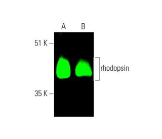
- rhodopsin Antibody (1D4) is a mouse monoclonal IgG1 κ rhodopsin antibody, cited in 77 publications, provided at 200 µg/ml
- raised against full length rhodopsin of bovine origin
- rhodopsin Antibody (1D4) is recommended for detection of rhodopsin of mouse, rat and human origin by WB, IP, IF, IHC(P) and ELISA; also reactive with additional species, including bovine
- Anti-rhodopsin Antibody (1D4) is available conjugated to agarose for IP; HRP for WB, IHC(P) and ELISA; and to either phycoerythrin or FITC for IF, IHC(P) and FCM
- also available conjugated to Alexa Fluor® 488, Alexa Fluor® 546, Alexa Fluor® 594 or Alexa Fluor® 647 for WB (RGB), IF, IHC(P) and FCM, and for use with RGB fluorescent imaging systems, such as iBright™ FL1000, FluorChem™, Typhoon, Azure and other comparable systems
- also available conjugated to Alexa Fluor® 680 or Alexa Fluor® 790 for WB (NIR), IF and FCM; for use with Near-Infrared (NIR) detection systems, such as LI-COR®Odyssey®, iBright™ FL1000, FluorChem™, Typhoon, Azure and other comparable systems
- m-IgG Fc BP-HRP, m-IgG1 BP-HRP and m-IgGκ BP-HRP are the preferred secondary detection reagents for rhodopsin Antibody (1D4) for WB and IHC(P) applications. These reagents are now offered in bundles with rhodopsin Antibody (1D4) (see ordering information below).

QUICK LINKS
SEE ALSO...
rhodopsin Antibody (1D4) is a mouse monoclonal IgG1 kappa light chain antibody that detects rhodopsin protein of mouse, rat, and human origin by western blotting (WB), immunoprecipitation (IP), immunofluorescence (IF), immunohistochemistry, and enzyme-linked immunosorbent assay (ELISA). anti-rhodopsin antibody (1D4) is available in both non-conjugated and various conjugated forms, including agarose, horseradish peroxidase (HRP), phycoerythrin (PE), fluorescein isothiocyanate (FITC), and multiple Alexa Fluor® conjugates. Rhodopsin, a member of the opsin subfamily of G protein-coupled receptors (GPCRs), plays a crucial role in the visual phototransduction pathway, allowing organisms to perceive light. Located in the outer segments of rod photoreceptor cells in the retina, rhodopsin is essential for vision in low-light conditions. The protein consists of a covalently bound chromophore, 11-cis-retinal, which undergoes isomerization upon photon absorption, triggering a series of conformational changes that activate G proteins and initiate the signaling cascade responsible for visual perception. This process enables the detection of light in dim environments and is vital for night vision and overall visual acuity. Mutations in the rhodopsin gene can lead to various forms of retinitis pigmentosa, a degenerative eye disease that can result in severe vision loss, highlighting rhodopsin′s importance in maintaining retinal health and function.
Alexa Fluor® is a trademark of Molecular Probes Inc., OR., USA
LI-COR® and Odyssey® are registered trademarks of LI-COR Biosciences
rhodopsin Antibody (1D4) References:
- Photoisomerization in rhodopsin. | Kandori, H., et al. 2001. Biochemistry (Mosc). 66: 1197-209. PMID: 11743865
- Rhodopsin-mediated retinitis pigmentosa. | Malanson, KM. and Lem, J. 2009. Prog Mol Biol Transl Sci. 88: 1-31. PMID: 20374723
- Phospholipid scrambling by rhodopsin. | Ernst, OP. and Menon, AK. 2015. Photochem Photobiol Sci. 14: 1922-31. PMID: 26179029
- Rhodopsin Oligomerization and Aggregation. | Park, PS. 2019. J Membr Biol. 252: 413-423. PMID: 31286171
- Flow of information in the light-triggered cyclic nucleotide cascade of vision. | Fung, BK., et al. 1981. Proc Natl Acad Sci U S A. 78: 152-6. PMID: 6264430
- The structure of bovine rhodopsin. | Hargrave, PA., et al. 1983. Biophys Struct Mech. 9: 235-44. PMID: 6342691
- G-protein diseases furnish a model for the turn-on switch. | Iiri, T., et al. 1998. Nature. 394: 35-8. PMID: 9665125
- Structure of rhodopsin. | Schertler, GF. 1998. Eye (Lond). 12 (Pt 3b): 504-10. PMID: 9775210
Ordering Information
| Product Name | Catalog # | UNIT | Price | Qty | FAVORITES | |
rhodopsin Antibody (1D4) | sc-57432 | 200 µg/ml | $322.00 | |||
rhodopsin Antibody (1D4): m-IgG Fc BP-HRP Bundle | sc-528623 | 200 µg Ab; 10 µg BP | $361.00 | |||
rhodopsin Antibody (1D4): m-IgGκ BP-HRP Bundle | sc-521049 | 200 µg Ab, 40 µg BP | $361.00 | |||
rhodopsin Antibody (1D4): m-IgG1 BP-HRP Bundle | sc-543035 | 200 µg Ab; 20 µg BP | $361.00 | |||
rhodopsin Antibody (1D4) AC | sc-57432 AC | 500 µg/ml, 25% agarose | $424.00 | |||
rhodopsin Antibody (1D4) HRP | sc-57432 HRP | 200 µg/ml | $322.00 | |||
rhodopsin Antibody (1D4) FITC | sc-57432 FITC | 200 µg/ml | $336.00 | |||
rhodopsin Antibody (1D4) PE | sc-57432 PE | 200 µg/ml | $349.00 | |||
rhodopsin Antibody (1D4) Alexa Fluor® 488 | sc-57432 AF488 | 200 µg/ml | $364.00 | |||
rhodopsin Antibody (1D4) Alexa Fluor® 546 | sc-57432 AF546 | 200 µg/ml | $364.00 | |||
rhodopsin Antibody (1D4) Alexa Fluor® 594 | sc-57432 AF594 | 200 µg/ml | $364.00 | |||
rhodopsin Antibody (1D4) Alexa Fluor® 647 | sc-57432 AF647 | 200 µg/ml | $364.00 | |||
rhodopsin Antibody (1D4) Alexa Fluor® 680 | sc-57432 AF680 | 200 µg/ml | $364.00 | |||
rhodopsin Antibody (1D4) Alexa Fluor® 790 | sc-57432 AF790 | 200 µg/ml | $364.00 |