")
pan CEA Antibody (H-8): sc-48364
- pan CEA Antibody (H-8) is a mouse monoclonal IgG1 κ pan CEA antibody, cited in 4 publications, provided at 200 µg/ml
- raised against amino acids 35-334 mapping near the N-terminus of CEA (carcinoembryonic antigen) of human origin
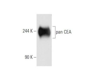
- pan CEA Antibody (H-8) is recommended for detection of pan CEA of human origin by WB, IP, IF, IHC(P) and ELISA
- Anti-pan CEA Antibody (H-8) is available conjugated to agarose for IP; HRP for WB, IHC(P) and ELISA; and to either phycoerythrin or FITC for IF, IHC(P) and FCM
- also available conjugated to Alexa Fluor® 488, Alexa Fluor® 546, Alexa Fluor® 594 or Alexa Fluor® 647 for WB (RGB), IF, IHC(P) and FCM, and for use with RGB fluorescent imaging systems, such as iBright™ FL1000, FluorChem™, Typhoon, Azure and other comparable systems
- also available conjugated to Alexa Fluor® 680 or Alexa Fluor® 790 for WB (NIR), IF and FCM; for use with Near-Infrared (NIR) detection systems, such as LI-COR®Odyssey®, iBright™ FL1000, FluorChem™, Typhoon, Azure and other comparable systems
- At present, we have not yet completed the identification of the preferred secondary detection reagent(s) for pan CEA Antibody (H-8). This work is in progress.

QUICK LINKS
SEE ALSO...
pan CEA Antibody (H-8) is a mouse monoclonal IgG1 kappa light chain antibody that detects pan CEA protein of human origin by western blotting (WB), immunoprecipitation (IP), immunofluorescence (IF), immunohistochemistry with paraffin-embedded sections (IHCP), and enzyme-linked immunosorbent assay (ELISA). anti-pan CEA antibody (H-8) is available in both non-conjugated and various conjugated forms, including agarose, horseradish peroxidase (HRP), phycoerythrin (PE), fluorescein isothiocyanate (FITC), and multiple Alexa Fluor® conjugates. Carcinoembryonic antigen (CEA) is a critical tumor marker widely utilized in serum immunoassays for carcinoma detection, playing a significant role in cancer diagnosis and monitoring. pan CEA (H-8) antibody targets proteins primarily located on the cell surface, where CEA is involved in cell adhesion processes essential for maintaining tissue architecture and facilitating communication between cells. This localization is particularly important in tumor biology, as alterations in cell adhesion can lead to increased tumor invasiveness and metastasis. CEACAM proteins, including pan CEA, are encoded by genes clustered on the long arm of chromosome 19, and expression levels can provide valuable insights into the progression of various malignancies.
Alexa Fluor® is a trademark of Molecular Probes Inc., OR., USA
LI-COR® and Odyssey® are registered trademarks of LI-COR Biosciences
pan CEA Antibody (H-8) References:
- Augmenting transgene expression from carcinoembryonic antigen (CEA) promoter via a GAL4 gene regulatory system. | Koch, PE., et al. 2001. Mol Ther. 3: 278-83. PMID: 11273768
- Keratin and carcinoembryonic antigen (CEA) in human melanoma cells. | Om, A., et al. 1991. Virchows Arch B Cell Pathol Incl Mol Pathol. 61: 81-7. PMID: 1720588
- Haplotypic diversity in human CEACAM genes: effects on susceptibility to meningococcal disease. | Callaghan, MJ., et al. 2008. Genes Immun. 9: 30-7. PMID: 17960155
- The CEACAM1 transmembrane domain, but not the cytoplasmic domain, directs internalization of human pathogens via membrane microdomains. | Muenzner, P., et al. 2008. Cell Microbiol. 10: 1074-92. PMID: 18081725
- The CEACAM1-mediated apoptosis pathway is activated by CEA and triggers dual cleavage of CEACAM1. | Nittka, S., et al. 2008. Oncogene. 27: 3721-8. PMID: 18278069
- CEACAM1 dynamics during neisseria gonorrhoeae suppression of CD4+ T lymphocyte activation. | Lee, HS., et al. 2008. J Immunol. 180: 6827-35. PMID: 18453603
- Altered splicing of CEACAM1 in breast cancer: identification of regulatory sequences that control splicing of CEACAM1 into long or short cytoplasmic domain isoforms. | Gaur, S., et al. 2008. Mol Cancer. 7: 46. PMID: 18507857
- CEACAM1, a SOX9 direct transcriptional target identified in the colon epithelium. | Zalzali, H., et al. 2008. Oncogene. 27: 7131-8. PMID: 18794798
- CEACAM1 inhibits Toll-like receptor 2-triggered antibacterial responses of human pulmonary epithelial cells. | Slevogt, H., et al. 2008. Nat Immunol. 9: 1270-8. PMID: 18836450
- Cytokine-induced CEACAM1 expression on keratinocytes is characteristic for psoriatic skin and contributes to a prolonged lifespan of neutrophils. | Rahmoun, M., et al. 2009. J Invest Dermatol. 129: 671-81. PMID: 18843289
- Interdependency of CEACAM-1, -3, -6, and -8 induced human neutrophil adhesion to endothelial cells. | Skubitz, KM. and Skubitz, AP. 2008. J Transl Med. 6: 78. PMID: 19077207
- Tissue demonstration of carcinoembryonic antigen (CEA) in ulcerative colitis. | Isaacson, P. 1976. Gut. 17: 561-7. PMID: 964689
Ordering Information
| Product Name | Catalog # | UNIT | Price | Qty | FAVORITES | |
pan CEA Antibody (H-8) | sc-48364 | 200 µg/ml | $322.00 | |||
pan CEA Antibody (H-8) AC | sc-48364 AC | 500 µg/ml, 25% agarose | $424.00 | |||
pan CEA Antibody (H-8) HRP | sc-48364 HRP | 200 µg/ml | $322.00 | |||
pan CEA Antibody (H-8) FITC | sc-48364 FITC | 200 µg/ml | $336.00 | |||
pan CEA Antibody (H-8) PE | sc-48364 PE | 200 µg/ml | $349.00 | |||
pan CEA Antibody (H-8) Alexa Fluor® 488 | sc-48364 AF488 | 200 µg/ml | $364.00 | |||
pan CEA Antibody (H-8) Alexa Fluor® 546 | sc-48364 AF546 | 200 µg/ml | $364.00 | |||
pan CEA Antibody (H-8) Alexa Fluor® 594 | sc-48364 AF594 | 200 µg/ml | $364.00 | |||
pan CEA Antibody (H-8) Alexa Fluor® 647 | sc-48364 AF647 | 200 µg/ml | $364.00 | |||
pan CEA Antibody (H-8) Alexa Fluor® 680 | sc-48364 AF680 | 200 µg/ml | $364.00 | |||
pan CEA Antibody (H-8) Alexa Fluor® 790 | sc-48364 AF790 | 200 µg/ml | $364.00 |