")
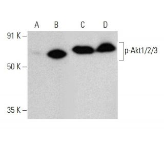
p-Akt1/2/3 Anticuerpo (B-5): sc-271966
- p-Akt1/2/3 Anticuerpo (B-5) es un monoclonal de ratón IgG2b κ, ver las 114 publicaciones, proporcionado como 200 µg/ml
- p-Akt1/2/3 Anticuerpo (B-5) es recomendado para detectar Thr 308 phosphorylated Akt1 and correspondingly Thr 309 phosphorylated Akt2 and correspondingly Thr 305 phosphorylated Akt3 de mouse, rat, human y origen, mediante WB, IP, IF, IHC(P) y ELISA; también reactivo con otras especies, incluyendo equine and avian
- p-Akt1/2/3 Anticuerpo (B-5) es disponible conjugado a agarosa para IP; HRP para WB, IHC(P) y ELISA; y tanto a phycoerythrin como a FITC para IF, IHC(P) y FCM
- también disponible conjugado a Alexa Fluor® 488, Alexa Fluor® 546, Alexa Fluor® 594 o Alexa Fluor® 647 para WB (RGB), IF, IHC (P) y FCM
- también disponible conjugado a Alexa Fluor® 680 o Alexa Fluor® 790 para WB (NIR), IF y FCM
- 2b BP-HRP">m-IgG2b BP-HRP es el reactivo de detección secundario preferido para p-Akt1/2/3 Anticuerpo (B-5) para aplicaciones WB e IHC(P). Este reactivo se ofrece ahora en un paquete con p-Akt1/2/3 Anticuerpo (B-5)(véase la información de pedido más abajo).

El anticuerpo p-Akt1/2/3 (B-5) es un anticuerpo monoclonal de ratón IgG2b κ que detecta la Akt1 fosforilada en Treonina 308 y correspondientemente la Akt2 fosforilada en Treonina 309 y la Akt3 fosforilada en Treonina 305 de origen humano, de rata y de ratón por WB, IP, IF, IHC(P) y ELISA. El anticuerpo p-Akt1/2/3 (B-5) está disponible en forma no conjugada, así como en múltiples formas conjugadas, incluyendo agarosa, HRP, PE, FITC y múltiples conjugados de Alexa Fluor®. La familia de quinasas serina/treonina Akt contiene varios miembros, incluyendo Akt1 (también designado como PKB o RacPK), Akt2 (también designado como PKBβ o RacPK-β) y Akt3 (también designado como PKBγ o protooncogén viral de timo 3), que muestran homología de secuencia con las familias de quinasa de proteína A y C y son codificados por el protooncogén c-Akt. Todos los miembros de la familia Akt tienen un dominio de homología de pleckstrin. Akt1 y Akt2 son activados por la estimulación de PDGF. Esta activación depende de los residuos de tirosina 740 y 751 de PDGFR-β, que se unen a la subunidad del complejo de fosfatidilinositol 3-quinasa (PI 3-quinasa). La activación de Akt1 por insulina o factor de crecimiento insulínico-1 (IGF-1) resulta en la fosforilación tanto de Treonina 308 como de Serina 473. Las proteínas Akt se fosforilan y activan en células estimuladas por insulina/IGF-1 por una quinasa(s) aguas arriba, y la activación de Akt1 y Akt2 es inhibida por el inhibidor de PI quinasa wortmannin. En conjunto, estos datos sugieren fuertemente que la proteína señala aguas abajo de las quinasas PI. Akt3 se fosforila en un residuo de serina en respuesta a la insulina. Sin embargo, la activación de Akt3 por insulina es inhibida por la activación previa de la proteína quinasa C a través de un mecanismo que no requiere la presencia del dominio PH. Akt3 se expresa en fibroblastos 3T3-L1, adipocitos y músculo esquelético y puede estar involucrado en varios procesos biológicos, incluyendo la diferenciación de adipocitos y músculo, síntesis de glucógeno, captación de glucosa, apoptosis y proliferación celular.
Alexa Fluor® es una marca registrada de Molecular Probes Inc., OR., USA
REIVEW LI-COR® y Odyssey® son marcas registradas de LI-COR Biosciences.
p-Akt1/2/3 Anticuerpo (B-5) Referencias:
- La expresión citoplasmática de Skp2 está asociada a p-Akt1 y predice un mal pronóstico en los carcinomas de mama humanos. | Liu, J., et al. 2012. PLoS One. 7: e52675. PMID: 23300741
- Descubrimiento de derivados de 4-(piperazin-1-il)-7H-pirrolo[2,3-d]pirimidina como inhibidores de Akt. | Liu, Y., et al. 2016. Arch Pharm (Weinheim). 349: 356-62. PMID: 26991997
- Slug inhibe la proliferación y la formación de tumores de células de cáncer de cuello de útero humano mediante la regulación al alza de las proteínas p21/p27 y la regulación a la baja de la actividad de la vía de señalización Wnt/β-catenina a través de la trans-supresión de la expresión de Akt1/p-Akt1. | Cui, N., et al. 2016. Oncotarget. 7: 26152-67. PMID: 27036045
- La quercetina inhibe la migración y la invasión de las células HCCLM3 mediante la supresión de la expresión de p-Akt1, MMP-2 y MMP-9 de la metaloproteinasa de matriz (MMP). | Lu, J., et al. 2018. Med Sci Monit. 24: 2583-2589. PMID: 29701200
- Las vías de señalización celular P-AKT2/SPK1 (P-SPK1) y P-MEK/P-ERK intervienen en la migración de macrófagos inducida por LPS. | Lei, Y., et al. 2019. Am J Transl Res. 11: 2725-2741. PMID: 31217849
- Expresión de Akt1 y p-Akt1 en subconjuntos de células T periféricas de pacientes con esclerosis múltiple. | Oktelik, FB., et al. 2021. Acta Neurol Belg. 121: 1777-1782. PMID: 33034831
- La Hexokinasa 2 promovió la motilidad celular y la metástasis a distancia mediante la elevación de la fibronectina a través de Akt1/p-Akt1 en células de cáncer cervical. | Chen, Q., et al. 2021. Cancer Cell Int. 21: 600. PMID: 34758823
- La hexoquinasa 2 promovió la motilidad y proliferación celular mediante la activación de Akt1/p-Akt1 en células de cáncer de ovario humano. | Tian, X., et al. 2022. J Ovarian Res. 15: 92. PMID: 35953860
- La importancia de p-AKT1 como marcador pronóstico y diana terapéutica en pacientes con cáncer de mama precoz positivo para receptores hormonales y receptor 2 del factor de crecimiento epidérmico humano. | Kim, JY., et al. 2022. J Breast Cancer. 25: 387-403. PMID: 36314765
- El eje IL-21/23 modula las citocinas inflamatorias y la expresión de RANKL en las células T CD4+ de la AR a través de la señalización p-Akt1. | Bhattacharya, G., et al. 2023. Front Immunol. 14: 1235514. PMID: 37809066
Información sobre pedidos
| Nombre del producto | Número de catálogo | UNIDAD | Precio | CANTIDAD | Favoritos | |
p-Akt1/2/3 Anticuerpo (B-5) | sc-271966 | 200 µg/ml | $322.00 | |||
Paquete de p-Akt1/2/3 (B-5): m-IgG2b BP-HRP | sc-548711 | 200 µg Ab; 10 µg BP | $361.00 | |||
p-Akt1/2/3 Anticuerpo (B-5) AC | sc-271966 AC | 500 µg/ml, 25% agarose | $424.00 | |||
p-Akt1/2/3 Anticuerpo (B-5) HRP | sc-271966 HRP | 200 µg/ml | $322.00 | |||
p-Akt1/2/3 Anticuerpo (B-5) FITC | sc-271966 FITC | 200 µg/ml | $336.00 | |||
p-Akt1/2/3 Anticuerpo (B-5) PE | sc-271966 PE | 200 µg/ml | $349.00 | |||
p-Akt1/2/3 Anticuerpo (B-5) Alexa Fluor® 488 | sc-271966 AF488 | 200 µg/ml | $364.00 | |||
p-Akt1/2/3 Anticuerpo (B-5) Alexa Fluor® 546 | sc-271966 AF546 | 200 µg/ml | $364.00 | |||
p-Akt1/2/3 Anticuerpo (B-5) Alexa Fluor® 594 | sc-271966 AF594 | 200 µg/ml | $364.00 | |||
p-Akt1/2/3 Anticuerpo (B-5) Alexa Fluor® 647 | sc-271966 AF647 | 200 µg/ml | $364.00 | |||
p-Akt1/2/3 Anticuerpo (B-5) Alexa Fluor® 680 | sc-271966 AF680 | 200 µg/ml | $364.00 | |||
p-Akt1/2/3 Anticuerpo (B-5) Alexa Fluor® 790 | sc-271966 AF790 | 200 µg/ml | $364.00 | |||
p-Akt1/2/3 (B-5) péptido neutralizante | sc-271966 P | 100 µg/0.5 ml | $69.00 |