
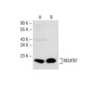
NDUFB7 Antibody (F-8): sc-365552
- NDUFB7 Antibody (F-8) is a mouse monoclonal IgG1 κ NDUFB7 antibody provided at 200 µg/ml
- raised against amino acids 1-137 representing full length NDUFB7 of human origin
- NDUFB7 Antibody (F-8) is recommended for detection of NDUFB7 of mouse, rat and human origin by WB, IP, IF and ELISA
- Anti-NDUFB7 Antibody (F-8) is available conjugated to agarose for IP; HRP for WB, IHC(P) and ELISA; and to either phycoerythrin or FITC for IF, IHC(P) and FCM
- also available conjugated to Alexa Fluor® 488, Alexa Fluor® 546, Alexa Fluor® 594 or Alexa Fluor® 647 for WB (RGB), IF, IHC(P) and FCM, and for use with RGB fluorescent imaging systems, such as iBright™ FL1000, FluorChem™, Typhoon, Azure and other comparable systems
- also available conjugated to Alexa Fluor® 680 or Alexa Fluor® 790 for WB (NIR), IF and FCM; for use with Near-Infrared (NIR) detection systems, such as LI-COR®Odyssey®, iBright™ FL1000, FluorChem™, Typhoon, Azure and other comparable systems
- m-IgG Fc BP-HRP and m-IgG1 BP-HRP are the preferred secondary detection reagents for NDUFB7 Antibody (F-8) for WB applications. These reagents are now offered in bundles with NDUFB7 Antibody (F-8) (see ordering information below).

NDUFB7 Antibody (F-8) is a mouse monoclonal IgG1 kappa light chain antibody that detects NDUFB7 protein of mouse, rat, and human origin by western blotting (WB), immunoprecipitation (IP), immunofluorescence (IF), and enzyme-linked immunosorbent assay (ELISA). NDUFB7 (F-8) antibody is available in both non-conjugated and various conjugated forms, including agarose, horseradish peroxidase (HRP), phycoerythrin (PE), fluorescein isothiocyanate (FITC), and multiple Alexa Fluor® conjugates. NDUFB7 protein, also known as NADH-ubiquinone oxioreductase B18 subunit, plays a crucial role as an accessory subunit of Complex I in the mitochondrial electron transport chain, which is essential for cellular respiration and energy production. This complex catalyzes the transfer of electrons from NADH to ubiquinone, facilitating the creation of an electrochemical proton gradient across the mitochondrial inner membrane, ultimately driving ATP synthesis. NDUFB7 protein is composed of 137 amino acids and is characterized by a sevenfold repeat of positively charged residues, suggesting involvement in protein-protein interactions that are vital for the stability and function of Complex I. Ubiquitously expressed, NDUFB7 is localized to the mitochondrial inner membrane on the matrix side, highlighting importance in mitochondrial function and energy metabolism.
Alexa Fluor® is a trademark of Molecular Probes Inc., OR., USA
LI-COR® and Odyssey® are registered trademarks of LI-COR Biosciences
NDUFB7 Antibody (F-8) References:
- Human mitochondrial complex I in health and disease. | Smeitink, J. and van den Heuvel, L. 1999. Am J Hum Genet. 64: 1505-10. PMID: 10330338
- Characterization of the human complex I NDUFB7 and 17.2-kDa cDNAs and mutational analysis of 19 genes of the HP fraction in complex I-deficient-patients. | Triepels, R., et al. 2000. Hum Genet. 106: 385-91. PMID: 10830904
- cDNA cloning of a novel cell adhesion protein expressed in human squamous carcinoma cells. | Wong, YC., et al. 1990. Biochem Biophys Res Commun. 166: 984-92. PMID: 2302251
- Intron based radiation hybrid mapping of 15 complex I genes of the human electron transport chain. | Emahazion, T., et al. 1998. Cytogenet Cell Genet. 82: 115-9. PMID: 9763677
- cDNA of eight nuclear encoded subunits of NADH:ubiquinone oxidoreductase: human complex I cDNA characterization completed. | Loeffen, JL., et al. 1998. Biochem Biophys Res Commun. 253: 415-22. PMID: 9878551
Ordering Information
| Product Name | Catalog # | UNIT | Price | Qty | FAVORITES | |
NDUFB7 Antibody (F-8) | sc-365552 | 200 µg/ml | $322.00 | |||
NDUFB7 Antibody (F-8): m-IgG Fc BP-HRP Bundle | sc-527379 | 200 µg Ab; 10 µg BP | $361.00 | |||
NDUFB7 Antibody (F-8): m-IgG1 BP-HRP Bundle | sc-532752 | 200 µg Ab; 20 µg BP | $361.00 | |||
NDUFB7 Antibody (F-8) AC | sc-365552 AC | 500 µg/ml, 25% agarose | $424.00 | |||
NDUFB7 Antibody (F-8) HRP | sc-365552 HRP | 200 µg/ml | $322.00 | |||
NDUFB7 Antibody (F-8) FITC | sc-365552 FITC | 200 µg/ml | $336.00 | |||
NDUFB7 Antibody (F-8) PE | sc-365552 PE | 200 µg/ml | $349.00 | |||
NDUFB7 Antibody (F-8) Alexa Fluor® 488 | sc-365552 AF488 | 200 µg/ml | $364.00 | |||
NDUFB7 Antibody (F-8) Alexa Fluor® 546 | sc-365552 AF546 | 200 µg/ml | $364.00 | |||
NDUFB7 Antibody (F-8) Alexa Fluor® 594 | sc-365552 AF594 | 200 µg/ml | $364.00 | |||
NDUFB7 Antibody (F-8) Alexa Fluor® 647 | sc-365552 AF647 | 200 µg/ml | $364.00 | |||
NDUFB7 Antibody (F-8) Alexa Fluor® 680 | sc-365552 AF680 | 200 µg/ml | $364.00 | |||
NDUFB7 Antibody (F-8) Alexa Fluor® 790 | sc-365552 AF790 | 200 µg/ml | $364.00 |