
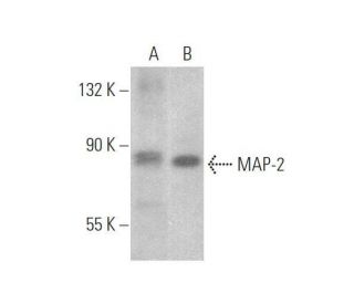
MAP-2 Antibody (AA6): sc-80013
- MAP-2 Antibody (AA6) is a mouse monoclonal IgG2a κ, cited in 5 publications, provided at 200 µg/ml
- raised against taxol-stabilized microtubules derived from brain of rat origin
- recommended for detection of MAP-2 of mouse, rat and human origin by WB, IP and IF
- See MAP-2 (A-4): sc-74421 for MAP-2 antibody conjugates, including AC, HRP, FITC, PE, Alexa Fluor® 488, 594, 647, 680 and 790.
- m-IgG Fc BP-HRP is the preferred secondary detection reagent for MAP-2 Antibody (AA6) for WB applications. This reagent is now offered in a bundle with MAP-2 Antibody (AA6) (see ordering information below).

MAP-2 Antibody (AA6) is a mouse monoclonal IgG2a antibody that detects microtubule-associated protein 2 (MAP-2) in mouse, rat, and human samples through applications such as western blotting (WB), immunoprecipitation (IP), and immunofluorescence (IF). MAP-2 plays a crucial role in the stabilization and assembly of microtubules, which are essential components of the cytoskeletal network that provide structural support and shape to cells. MAP-2 (AA6) antibody targets proteins primarily located in neuronal cell bodies and dendrites, where MAP-2 contributes to the organization of the microtubule network, facilitating intracellular transport and maintaining the integrity of neuronal architecture. The proper functioning of MAP-2 is vital for neuronal development and plasticity, as MAP-2 influences the growth and branching of dendrites, which are critical for synaptic connectivity and communication between neurons. Dysregulation of MAP-2 has been associated with various neurodegenerative diseases, making MAP-2 an important target for research in understanding neuronal health and disease mechanisms. Anti-MAP-2 antibody (AA6) is an invaluable tool for researchers studying the dynamics of microtubules and their associated proteins in various biological contexts.
Alexa Fluor® is a trademark of Molecular Probes Inc., OR., USA
LI-COR® and Odyssey® are registered trademarks of LI-COR Biosciences
MAP-2 Antibody (AA6) References:
- Immunocytochemical expression of microtubule-associated protein-2 (MAP-2) in small cell lung cancer cell lines with neuronal-like processes. | Tanaka, K. and Terasaki, T. 2002. Jpn J Cancer Res. 93: 699-705. PMID: 12079519
- Dietary aluminum selectively decreases MAP-2 in brains of developing and adult rats. | Johnson, GV., et al. 1992. Neurotoxicology. 13: 463-74. PMID: 1331920
- The susceptibility of MAP-2 to proteolytic degradation increases when bound to tubulin. | Grau, E., et al. 1992. Neurochem Res. 17: 967-71. PMID: 1508306
- Altered expression of MAP-2, GAP-43, and synaptophysin in the hippocampus of rats with chronic cerebral hypoperfusion correlates with cognitive impairment. | Liu, HX., et al. 2005. Brain Res Mol Brain Res. 139: 169-77. PMID: 15964096
- Motor Skill Training Promotes Sensorimotor Recovery and Increases Microtubule-Associated Protein-2 (MAP-2) Immunoreactivity in the Motor Cortex after Intracerebral Hemorrhage in the Rat. | Santos, MV., et al. 2013. ISRN Neurol. 2013: 159184. PMID: 23956876
- Anti-200 kDa neurofilament antibody cross-reacts with microtubule-associated protein-2 (MAP-2). | Allende, ML., et al. 1989. J Neurosci Res. 22: 130-3. PMID: 2468783
- Expression of MAP 2 by haemangioblastomas: an immunohistochemical study with implications for diagnosis. | Malafronte, PJ., et al. 2014. Pathology. 46: 450-1. PMID: 24977729
- Antibodies to microtubule-associated protein-2 in the cerebrospinal fluid are a useful diagnostic biomarker for neuropsychiatric systemic lupus erythematosus. | Yamada, Y., et al. 2016. Mod Rheumatol. 26: 562-8. PMID: 26667376
- Ammonium injection induces an N-methyl-D-aspartate receptor-mediated proteolysis of the microtubule-associated protein MAP-2. | Felipo, V., et al. 1993. J Neurochem. 60: 1626-30. PMID: 8473887
- Disruption of MAP-2 immunostaining in rat hippocampus after traumatic brain injury. | Folkerts, MM., et al. 1998. J Neurotrauma. 15: 349-63. PMID: 9605349
Ordering Information
| Product Name | Catalog # | UNIT | Price | Qty | FAVORITES | |
MAP-2 Antibody (AA6) | sc-80013 | 200 µg/ml | $322.00 | |||
MAP-2 Antibody (AA6): m-IgG Fc BP-HRP Bundle | sc-539555 | 200 µg Ab; 10 µg BP | $361.00 |