")
: sc-166245. Análisis por Western blot de la expresión de HAP1 en lisado de células enteras COLO 320DM.")
: sc-166245. Tinción de inmunoperoxidasa de tejido de corteza cerebral humano fijado con formalina e incluido en parafina que muestra tinción citoplasmática de células neuronales y gliales.")
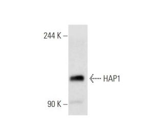
: sc-166245. Análisis por Western blot de la expresión de HAP1 en lisados de células enteras Neuro-2A.")
HAP1 Anticuerpo (C-3): sc-166245
- HAP1 Anticuerpo C-3 es un monoclonal de ratón IgG1 κ HAP1 Anticuerpo, ver las 1 publicaciones, proporcionado como 200 µg/ml
- planteada frente a los aminoácidos 329-628 que corresponden al C-terminus de HAP1B de mouse origen
- HAP1 Anticuerpo (C-3) es recomendado para detectar HAP1 de mouse, rat y human origen, mediante WB, IP, IF, IHC(P) y ELISA
- HAP1 Anticuerpo (C-3) es disponible conjugado a agarosa para IP; HRP para WB, IHC(P) y ELISA; y tanto a phycoerythrin como a FITC para IF, IHC(P) y FCM
- también disponible conjugado a Alexa Fluor® 488, Alexa Fluor® 546, Alexa Fluor® 594 o Alexa Fluor® 647 para WB (RGB), IF, IHC (P) y FCM
- también disponible conjugado a Alexa Fluor® 680 o Alexa Fluor® 790 para WB (NIR), IF y FCM
- m-IgG Fc BP-HRP y m-IgG1 BP-HRP son los reactivos de detección secundarios preferidos para HAP1 Anticuerpo (C-3) for WB and IHC(P) applications. Estos reactivos se ofrecen ahora en paquetes con HAP1 Anticuerpo (C-3)(véase la información de pedido más abajo).

ENLACES RÁPIDOS
El anticuerpo HAP1 (C-3) es un anticuerpo monoclonal de ratón IgG1 κ HAP1 (también designado como anticuerpo HAP1) que detecta la proteína HAP1 de origen ratón, rata y humano por WB, IP, IF, IHC(P) y ELISA. El anticuerpo HAP1 (C-3) está disponible tanto en forma de anticuerpo anti-HAP1 no conjugado, como en múltiples formas conjugadas de anticuerpo anti-HAP1, incluyendo agarosa, HRP, PE, FITC y múltiples conjugados de Alexa Fluor®. HAP1 (proteína asociada a huntingtina 1) se une a huntingtina. La huntingtina es una proteína que contiene una región de poliglutamina y cuando el número de repeticiones de glutamina excede 35, el gen codifica una versión de huntingtina que conduce a la enfermedad de Huntington (HD). La capacidad de HAP1 para unirse a huntingtina se ve potenciada por una región expandida de repeticiones de poliglutamina. HAP1 muestra localización neuronal y se mueve con huntingtina en las fibras nerviosas. HAP1 se expresa principalmente en tejido cerebral, con una mayor expresión en el bulbo olfatorio y el tronco cerebral. En la rata, se ha demostrado que HAP1 se asocia con varios orgánulos intracelulares. En el ratón, HAP1 se localiza en orgánulos de membrana, incluyendo endosomas grandes, estructuras tubulovesiculares y vesículas en desarrollo en neuronas.
Alexa Fluor® es una marca registrada de Molecular Probes Inc., OR., USA
REIVEW LI-COR® y Odyssey® son marcas registradas de LI-COR Biosciences.
HAP1 Anticuerpo (C-3) Referencias:
- Agregados nucleares y neuropilares en la enfermedad de Huntington: relación con la neuropatología. | Gutekunst, CA., et al. 1999. J Neurosci. 19: 2522-34. PMID: 10087066
- Una proteína asociada a la huntingtina enriquecida en el cerebro con implicaciones para la patología. | Li, XJ., et al. 1995. Nature. 378: 398-402. PMID: 7477378
- Un nuevo gen que contiene una repetición de trinucleótidos expandida e inestable en los cromosomas de la enfermedad de Huntington. Grupo de Investigación Colaborativa sobre la Enfermedad de Huntington. | . 1993. Cell. 72: 971-83. PMID: 8458085
- Proteína asociada a la huntingtina (HAP1): su localización neuronal en el cerebro se asemeja a la de la óxido nítrico sintasa neuronal. | Li, XJ., et al. 1996. Proc Natl Acad Sci U S A. 93: 4839-44. PMID: 8643490
- Transporte rápido y movimiento retrógrado de huntingtina y HAP 1 en axones. | Block-Galarza, J., et al. 1997. Neuroreport. 8: 2247-51. PMID: 9243620
- Enfermedad de Huntington. | Gusella, JF., et al. 1996. Cold Spring Harb Symp Quant Biol. 61: 615-26. PMID: 9246488
- Análisis de la proteína 1 asociada a Huntingtina en cerebro de ratón y neuronas estriatales inmortalizadas. | Martin, EJ., et al. 1999. J Comp Neurol. 403: 421-30. PMID: 9888310
Información sobre pedidos
| Nombre del producto | Número de catálogo | UNIDAD | Precio | CANTIDAD | Favoritos | |
HAP1 Anticuerpo (C-3) | sc-166245 | 200 µg/ml | $322.00 | |||
Paquete de HAP1 (C-3): m-IgG Fc BP-HRP | sc-527147 | 200 µg Ab; 10 µg BP | $361.00 | |||
Paquete de HAP1 (C-3): m-IgG1 BP-HRP | sc-532520 | 200 µg Ab; 20 µg BP | $361.00 | |||
HAP1 Anticuerpo (C-3) AC | sc-166245 AC | 500 µg/ml, 25% agarose | $424.00 | |||
HAP1 Anticuerpo (C-3) HRP | sc-166245 HRP | 200 µg/ml | $322.00 | |||
HAP1 Anticuerpo (C-3) FITC | sc-166245 FITC | 200 µg/ml | $336.00 | |||
HAP1 Anticuerpo (C-3) PE | sc-166245 PE | 200 µg/ml | $349.00 | |||
HAP1 Anticuerpo (C-3) Alexa Fluor® 488 | sc-166245 AF488 | 200 µg/ml | $364.00 | |||
HAP1 Anticuerpo (C-3) Alexa Fluor® 546 | sc-166245 AF546 | 200 µg/ml | $364.00 | |||
HAP1 Anticuerpo (C-3) Alexa Fluor® 594 | sc-166245 AF594 | 200 µg/ml | $364.00 | |||
HAP1 Anticuerpo (C-3) Alexa Fluor® 647 | sc-166245 AF647 | 200 µg/ml | $364.00 | |||
HAP1 Anticuerpo (C-3) Alexa Fluor® 680 | sc-166245 AF680 | 200 µg/ml | $364.00 | |||
HAP1 Anticuerpo (C-3) Alexa Fluor® 790 | sc-166245 AF790 | 200 µg/ml | $364.00 |