")
FAK Anticuerpo (H-1): sc-1688
- FAK Anticuerpo (H-1) es un monoclonal de ratón IgG1 κ, ver las 111 publicaciones, proporcionado como 200 µg/ml
- producido contra los amino ácidos 903-1052 de FAK de origen mouse
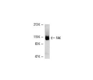
- FAK Anticuerpo (H-1) es recomendado para detectar FAK p125 de mouse, rat y human origen, mediante WB, IP, IF, IHC(P) y FCM
- FAK Anticuerpo (H-1) es disponible conjugado a ficoeritrina para IF, IHC(P) y FCM
- también disponible conjugado a Alexa Fluor® 594 para IF, IHC(P) y FCM
- también disponible conjugado a Alexa Fluor® 680 o Alexa Fluor® 790 para WB (NIR), IF y FCM
- m-IgG Fc BP-HRP y m-IgG1 BP-HRP son los reactivos de detección secundarios preferidos para FAK Anticuerpo (H-1) for WB and IHC(P) applications. Estos reactivos se ofrecen ahora en paquetes con FAK Anticuerpo (H-1)(véase la información de pedido más abajo).

ENLACES RÁPIDOS
VER TAMBIÉN ....
El anticuerpo FAK (H-1) es un anticuerpo monoclonal de ratón IgG1 κ que detecta FAK p125 de origen ratón, rata y humano por WB, IP, IF, IHC(P) y FCM. El anticuerpo FAK (H-1) está disponible en formas conjugadas y no conjugadas. La quinasa de adhesión focal fue identificada inicialmente como un sustrato importante para la actividad intrínseca de tirosina quinasa de la proteína Src codificada por pp60. La secuencia de aminoácidos deducida de FAK p125 ha demostrado que es una quinasa de tirosina citoplasmática cuya secuencia y organización estructural son únicas en comparación con otras proteínas descritas hasta la fecha. La localización de p125 por inmunofluorescencia sugiere que se encuentra principalmente en las adhesiones focales celulares, lo que lleva a su designación como quinasa de adhesión focal (FAK). FAK se concentra en el borde basal solo de aquellas queratinocitos basales que están migrando activamente y proliferando rápidamente en la reparación de quemaduras, y se activa y localiza en las adhesiones focales de queratinocitos en cultivo. Por lo tanto, se ha postulado que FAK puede tener un papel importante in vivo en la reepitelización de heridas humanas. También se ha demostrado que la actividad de la quinasa de tirosina de la proteína FAK aumenta en células estimuladas para crecer mediante el uso de neuropéptidos mitogénicos o neurotransmisores que actúan a través de receptores acoplados a proteínas G.
Alexa Fluor® es una marca registrada de Molecular Probes Inc., OR., USA
REIVEW LI-COR® y Odyssey® son marcas registradas de LI-COR Biosciences.
FAK Anticuerpo (H-1) Referencias:
- La activación de los receptores transformantes acoplados a proteínas G induce la rápida fosforilación en tirosina de proteínas celulares, entre ellas la p125FAK y el sustrato p130 v-src. | Gutkind, JS. and Robbins, KC. 1992. Biochem Biophys Res Commun. 188: 155-61. PMID: 1329743
- Regulación de la proteína tirosina cinasa asociada a la adhesión focal tanto por la adhesión celular como por la transformación oncogénica. | Guan, JL. and Shalloway, D. 1992. Nature. 358: 690-2. PMID: 1379699
- Estimulación de la fosforilación de tirosina por la bombesina, la vasopresina y la endotelina en células 3T3 suizas. Identificación de una nueva tirosina quinasa como sustrato principal. | Zachary, I., et al. 1992. J Biol Chem. 267: 19031-4. PMID: 1382065
- Fosforilación dependiente de integrina y activación de la proteína tirosina quinasa pp125FAK en plaquetas. | Lipfert, L., et al. 1992. J Cell Biol. 119: 905-12. PMID: 1385445
- Proteína tirosina cinasa de adhesión focal fosforilada en respuesta a la adhesión celular a la fibronectina. | Hanks, SK., et al. 1992. Proc Natl Acad Sci U S A. 89: 8487-91. PMID: 1528852
- pp125FAK una proteína-tirosina quinasa estructuralmente distintiva asociada a las adhesiones focales. | Schaller, MD., et al. 1992. Proc Natl Acad Sci U S A. 89: 5192-6. PMID: 1594631
- La autofosforilación de la quinasa de adhesión focal, pp125FAK, dirige la unión dependiente de SH2 de pp60src. | Schaller, MD., et al. 1994. Mol Cell Biol. 14: 1680-8. PMID: 7509446
- Papel potencial de la quinasa de adhesión focal en queratinocitos en migración y proliferación cerca de heridas epidérmicas y en cultivo. | Gates, RE., et al. 1994. Cell Growth Differ. 5: 891-9. PMID: 7986754
Información sobre pedidos
| Nombre del producto | Número de catálogo | UNIDAD | Precio | CANTIDAD | Favoritos | |
FAK Anticuerpo (H-1) | sc-1688 | 200 µg/ml | $322.00 | |||
Paquete de FAK (H-1): m-IgG Fc BP-HRP | sc-526472 | 200 µg Ab; 10 µg BP | $361.00 | |||
Paquete de FAK (H-1): m-IgG1 BP-HRP | sc-531845 | 200 µg Ab; 20 µg BP | $361.00 | |||
FAK Anticuerpo (H-1) PE | sc-1688 PE | 200 µg/ml | $349.00 | |||
FAK Anticuerpo (H-1) Alexa Fluor® 546 | sc-1688 AF546 | 200 µg/ml | $364.00 | |||
FAK Anticuerpo (H-1) Alexa Fluor® 594 | sc-1688 AF594 | 200 µg/ml | $364.00 | |||
FAK Anticuerpo (H-1) Alexa Fluor® 680 | sc-1688 AF680 | 200 µg/ml | $364.00 | |||
FAK Anticuerpo (H-1) Alexa Fluor® 790 | sc-1688 AF790 | 200 µg/ml | $364.00 |