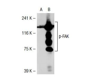
Date published: 2025-9-9

00800 4573 8000

SCBT Portrait Logo
Seach Input

p-FAK Antikörper (2D11): sc-81493

4.1(23)
Produkt bewertenBitte stellen Sie eine Frage

Datenblätter
  • p-FAK Antikörper (2D11) ist ein Maus monoklonales IgG1 κ, verwendet in 61 wissenschaftlichen Veröffentlichungen, in einer Menge von 50 µg/0.5 ml
  • gegen Phosphopeptid, das den Aminosäureresten entspricht, die Tyrosin 397 von FAK mit Ursprung in human umgeben,
  • Empfohlen für die Detektion von Tyr 397 phosphorylated FAK aus der Spezies mouse, rat, human und canine per WB und IP
  • Bitte kontaktieren Sie uns um ein 10 µg GRATIS Sample vom p-FAK (2D11): sc-81493 zu erhalten.
  • 1 BP-HRP">m-IgG1 BP-HRP und m-IgGκ BP-HRP sind die bevorzugten sekundären Nachweisreagenzien für p-FAK Antikörper (2D11) für WB-Anwendungen. Diese Reagenzien werden jetzt in Bündeln mit p-FAK Antikörper (2D11) angeboten(siehe Bestellinformationen unten).

Direktverknüpfungen

Siehe auch...

Der p-FAK-Antikörper (2D11) ist ein monoklonaler Maus IgG1 κ p-FAK-Antikörper, der Tyr 397 phosphorylierte FAK von Maus-, Ratte- und menschlicher Herkunft mittels WB und IP detektiert. Der p-FAK-Antikörper (2D11) ist als nicht-konjugierte Form des Anti-p-FAK-Antikörpers erhältlich. Die Aktivierung von Integrinen in der extrazellulären Matrix (ECM) eukaryotischer Zellen fördert die Bildung von Membranadherenskomplexen, die als Fokaladhesionen bekannt sind und die zytoskelettale Proteine und Protein-Tyrosinkinasen wie die Fokaladhesionskinase (FAK) umfassen können. Phosphorylierungsereignisse in Fokaladhesionen beeinflussen zahlreiche Prozesse, darunter mitogene Signalisierung, Zellüberleben und Zellmigration. FAK ist eine nicht-Rezeptor-Tyrosinkinase, die ubiquitär exprimiert und zwischen Arten hoch konserviert ist. FAK wird von Integrin-Clustern rekrutiert und je nach den vorhandenen Effektormolekülen in der Fokaladhesion variabel phosphoryliert. Die Phosphorylierung von FAK Tyr 397 nimmt während der Serumstarre, Kontaktinhibition und Zellzyklusarrest ab, Bedingungen, unter denen die aktivierende Phosphorylierung von FAK Tyr 407 zunimmt.

Ausschließlich für Forschungszwecke. Nicht für diagnostische oder therapeutische Zwecke bestimmt.

Alexa Fluor® ist ein Markenzeichen von Molecular Probes Inc., OR., USA

LI-COR® und Odyssey® sind Markenzeichen von LI-COR Biosciences

p-FAK Antikörper (2D11) Literaturhinweise:

  1. Regulierung der fokalen Adhäsions-assoziierten Protein-Tyrosin-Kinase durch zelluläre Adhäsion und onkogene Transformation.  |  Guan, JL. and Shalloway, D. 1992. Nature. 358: 690-2. PMID: 1379699
  2. Integrin-abhängige Phosphorylierung und Aktivierung der Protein-Tyrosin-Kinase pp125FAK in Blutplättchen.  |  Lipfert, L., et al. 1992. J Cell Biol. 119: 905-12. PMID: 1385445
  3. Fokale Adhäsionsprotein-Tyrosinkinase, die als Reaktion auf die Zellbindung an Fibronektin phosphoryliert wird.  |  Hanks, SK., et al. 1992. Proc Natl Acad Sci U S A. 89: 8487-91. PMID: 1528852
  4. pp125FAK, eine strukturell charakteristische Protein-Tyrosin-Kinase, die mit fokalen Adhäsionen assoziiert ist.  |  Schaller, MD., et al. 1992. Proc Natl Acad Sci U S A. 89: 5192-6. PMID: 1594631
  5. Die Focal Adhesion Kinase wird durch Phosphorylierung an Tyrosin 407 negativ reguliert.  |  Lim, Y., et al. 2007. J Biol Chem. 282: 10398-404. PMID: 17303567
  6. mTORC1/rpS6- und p-FAK-Y407-Signalübertragung reguliert die Spermatogenese: Erkenntnisse aus Studien zum Adudin-Pharmazeutika/Toxikum-Modell.  |  Wang, L., et al. 2022. Semin Cell Dev Biol. 121: 53-62. PMID: 33867214
  7. Phosphoryliertes Paxillin und phosphoryliertes FAK bilden Subregionen innerhalb fokaler Adhäsionen.  |  Bachmann, M., et al. 2022. J Cell Sci. 135: PMID: 35343568
  8. Von karzinomassoziierten Fibroblasten stammende lysyloxidase-reiche extrazelluläre Vesikel vermitteln die Kollagenvernetzung und fördern die epithelial-mesenchymale Transition über den p-FAK/p-paxillin/YAP-Signalweg.  |  Liu, X., et al. 2023. Int J Oral Sci. 15: 32. PMID: 37532712
  9. Die Autophosphorylierung der fokalen Adhäsionskinase, pp125FAK, steuert die SH2-abhängige Bindung von pp60src.  |  Schaller, MD., et al. 1994. Mol Cell Biol. 14: 1680-8. PMID: 7509446
  10. Nachweis der In-vivo-Phosphorylierung der Grb2-SH2-Domänen-Bindungsstelle an der fokalen Adhäsionskinase durch Protein-Tyrosin-Kinasen der Src-Familie.  |  Schlaepfer, DD. and Hunter, T. 1996. Mol Cell Biol. 16: 5623-33. PMID: 8816475

Bestellinformation

ProduktKatalog #EINHEITPreisANZAHLFavoriten

p-FAK Antikörper (2D11)

sc-81493
50 µg/0.5 ml
$316.00

p-FAK (2D11): m-IgGκ BP-HRP Bundle

sc-534469
50 µg Ab; 40 µg BP
$354.00

p-FAK (2D11): m-IgG1 BP-HRP Bundle

sc-538867
50 µg Ab; 20 µg BP
$354.00

How the dilution of this antibody should be prepared for use in western blotting

Gefragt von: Anonym
Thank you for your question. p-FAK (2D11) is recommended for detection of Tyr 397 phosphorylated FAK of mouse, rat, human and canine origin by Western Blotting with a starting dilution of 1:200, and a dilution range of 1:100-1:1000.
Beantwortet von: Tech Support Europe
Veröffentlichungsdatum: 2025-01-21

What is the best secondary antibody for this mouse monoclonal?

Gefragt von: Anna99
We recommend using one our exclusive Mouse IgG Binding Proteins as a secondary detection reagent. A complete list of available binding proteins is available on our website here: https://www.scbt.com/scbt/browse/support-products-mouse-igg-binding-proteins/_/N-ecrety
Beantwortet von: Technical Service 11
Veröffentlichungsdatum: 2017-01-13

I am using the LiCor/Odyssey system. Which secondary antibody do you recommend using with p-FAK Antibody (2D11): sc-81493 ?

Gefragt von: Anna99
We recommend using one of our our exclusive Mouse IgG Binding Proteins (m-IgGκ) conjugated to CFL 680 or CFL 790. They are suitable for use with Near-Infrared (NIR) detection systems, such as LI-COR/Odyssey and other comparable systems. A complete list of available binding proteins is available on our website here: https://www.scbt.com/scbt/browse/support-products-mouse-igg-binding-proteins/_/N-ecrety
Beantwortet von: Technical Service 11
Veröffentlichungsdatum: 2017-01-13
  • y_2025, m_8, d_29, h_8CST
  • bvseo_bulk, prod_bvqa, vn_bulk_3.0.42
  • cp_1, bvpage1
  • co_hasquestionsanswers, tq_3
  • loc_de_DE, sid_81493, prod, sort_[SortEntry(order=LAST_APPROVED_ANSWER_SUBMISSION_TIME, direction=DESCENDING)]
  • clientName_scbt
  • bvseo_sdk, java_sdk, bvseo-4.0.0
  • CLOUD, getContent, 119ms
  • QUESTIONS, PRODUCT
Rated 4 von 5 von aus Works well in WBThis antibody works well in WB, I used it to detect p-FAK in Huh7, dil:1:500
Veröffentlichungsdatum: 2020-02-21
Rated 1 von 5 von aus Not working antibodyI recived a free sample from this antibody and it din´t work. I am working with MDCK cells also I charge a sample from HeLa as a positive control, I used 1:50, 1:200 and 1:500 dilution and in any of those I saw a band on the western blot. I used GAPDH as a charge control and I tested my secondary antibody as well with no failures.
Veröffentlichungsdatum: 2019-07-22
Rated 2 von 5 von aus Non-specific bands obscure resultsI tried this on lysates from both mouse and humans cells in experiments where I was blotting for several other proteins at the same time. The others worked, but there was a strong band too large to be P-FAK that made it very difficult to get a result. The FAK (B-8) antibody worked to confirm that this larger band was not FAK. I suppose you could get a result if you ran a low % gel for a long time to get more separation.
Veröffentlichungsdatum: 2019-01-18
Rated 5 von 5 von aus good band in WBWorks good in WB with HCECs human corneal epithelial cells .
Veröffentlichungsdatum: 2018-11-22
Rated 5 von 5 von aus Nice band in WBp-FAK had strong band in WB in human leukemia cell line Molt4
Veröffentlichungsdatum: 2018-07-09
Rated 5 von 5 von aus Good for WB in cell lysatesWB using p-FAK (2D11) 1:1000 in 76NE6 cells, clear band detected.
Veröffentlichungsdatum: 2018-05-21
Rated 5 von 5 von aus Fantastic for Western BlotUsed this antibody at 1:1000 dilution on a western blot with 100 ug of protein from tissue and another blot with 60 ug of protein from cancer cell lines. Both blots came out absolutely crystal clear.
Veröffentlichungsdatum: 2018-02-21
Rated 5 von 5 von aus Good antibody!1:500 diluted for wb of rat protein,the band is quite strong and special!
Veröffentlichungsdatum: 2017-12-30
  • y_2025, m_8, d_29, h_8
  • bvseo_bulk, prod_bvrr, vn_bulk_3.0.42
  • cp_1, bvpage1
  • co_hasreviews, tv_0, tr_23
  • loc_de_DE, sid_81493, prod, sort_[SortEntry(order=SUBMISSION_TIME, direction=DESCENDING)]
  • clientName_scbt
  • bvseo_sdk, java_sdk, bvseo-4.0.0
  • CLOUD, getReviews, 8ms
  • REVIEWS, PRODUCT
p-FAK Antikörper (2D11) wurde bewertet mit 4.1 von 5 von 23.
  • y_2025, m_8, d_29, h_8
  • bvseo_bulk, prod_bvrr, vn_bulk_3.0.42
  • cp_1, bvpage1
  • co_hasreviews, tv_0, tr_23
  • loc_de_DE, sid_81493, prod, sort_[SortEntry(order=SUBMISSION_TIME, direction=DESCENDING)]
  • clientName_scbt
  • bvseo_sdk, java_sdk, bvseo-4.0.0
  • CLOUD, getAggregateRating, 127ms
  • REVIEWS, PRODUCT