
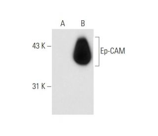
Ep-CAM Antibody (3H2141): sc-71056
- Ep-CAM Antibody (3H2141) is a mouse monoclonal IgG1 κ provided at 200 µg/ml
- raised against HT29 carcinoma cell line of human origin
- recommended for detection of Ep-CAM of human origin by WB, IP, IF and IHC(P)
- See Ep-CAM (C-10): sc-25308 for Ep-CAM antibody conjugates, including AC, HRP, FITC, PE, Alexa Fluor® 488, 594, 647, 680 and 790.
- m-IgG Fc BP-HRP and m-IgG1 BP-HRP are the preferred secondary detection reagents for Ep-CAM Antibody (3H2141) for WB and IHC(P) applications. These reagents are now offered in bundles with Ep-CAM Antibody (3H2141) (see ordering information below).

QUICK LINKS
SEE ALSO...
Ep-CAM Antibody (3H2141) is a mouse monoclonal IgG1 antibody that detects Ep-CAM in human samples through various applications including western blotting (WB), immunoprecipitation (IP), immunofluorescence (IF), and immunohistochemistry. Ep-CAM, also known as tumor-associated calcium signal transducer 1 and MK-1, is a monomeric membrane glycoprotein predominantly expressed in normal human epithelial tissues and various carcinomas, making Ep-CAM a significant marker for cancer research. The human Ep-CAM gene encodes a 314 amino acid protein that exists in two forms, with the major form being more prevalent and the minor form being reduced upon treatment with the amino-glycosylation inhibitor tunicamycin. Notably, Ep-CAM is characterized by an extracellular domain, which contains two epidermal growth factor-like repeats followed by a cysteine-poor region; this unique structure is crucial for adhesion properties, facilitating cell-cell interactions that are vital for maintaining tissue integrity. Given Ep-CAM′s overexpression in numerous carcinomas, anti-Ep-CAM antibody (3H2141) serves as a promising tool for both visualization and therapeutic intervention of human solid tumors, underscoring Ep-CAM′s importance in cancer diagnostics and treatment strategies.
Alexa Fluor® is a trademark of Molecular Probes Inc., OR., USA
LI-COR® and Odyssey® are registered trademarks of LI-COR Biosciences
Ep-CAM Antibody (3H2141) References:
- A relevant immunomagnetic assay to detect and characterize epithelial cell adhesion molecule-positive cells in bone marrow from patients with breast carcinoma: immunomagnetic purification of micrometastases. | Choesmel, V., et al. 2004. Cancer. 101: 693-703. PMID: 15305398
- Expression of epithelial cell adhesion molecule in gallbladder carcinoma and its correlation with clinicopathologic variables. | Prince, S., et al. 2008. Am J Clin Pathol. 129: 424-9. PMID: 18285265
- Oncostatin m renders epithelial cell adhesion molecule-positive liver cancer stem cells sensitive to 5-Fluorouracil by inducing hepatocytic differentiation. | Yamashita, T., et al. 2010. Cancer Res. 70: 4687-97. PMID: 20484035
- Selection of DNA aptamers against epithelial cell adhesion molecule for cancer cell imaging and circulating tumor cell capture. | Song, Y., et al. 2013. Anal Chem. 85: 4141-9. PMID: 23480100
- Epithelial cell adhesion molecule-positive circulating tumor cells as predictive biomarker in patients with prostate cancer. | Amato, RJ., et al. 2013. Urology. 81: 1303-7. PMID: 23622774
- Circulating tumor cell enumeration with a combination of epithelial cell adhesion molecule- and cell-surface vimentin-based methods for monitoring breast cancer therapeutic response. | Satelli, A., et al. 2015. Clin Chem. 61: 259-66. PMID: 25336717
- Immunohistochemical expression of epithelial cell adhesion molecule (EpCAM) in mucoepidermoid carcinoma compared to normal salivary gland tissues. | Kamal, NM., et al. 2017. Arch Oral Biol. 79: 87-94. PMID: 28347886
- Ultrasensitive amperometric aptasensor for the epithelial cell adhesion molecule by using target-driven toehold-mediated DNA recycling amplification. | Chen, Q., et al. 2018. Mikrochim Acta. 185: 202. PMID: 29594643
- Wash-free and selective imaging of epithelial cell adhesion molecule (EpCAM) expressing cells with fluorogenic peptide ligands. | K C, TB., et al. 2018. Biochem Biophys Res Commun. 500: 283-287. PMID: 29660346
- A meta-analysis and The Cancer Genome Atlas data of prostate cancer risk and prognosis using epithelial cell adhesion molecule (EpCAM) expression. | Hu, Y., et al. 2019. BMC Urol. 19: 67. PMID: 31324239
Ordering Information
| Product Name | Catalog # | UNIT | Price | Qty | FAVORITES | |
Ep-CAM Antibody (3H2141) | sc-71056 | 200 µg/ml | $322.00 | |||
Ep-CAM Antibody (3H2141): m-IgG Fc BP-HRP Bundle | sc-539339 | 200 µg Ab; 10 µg BP | $361.00 | |||
Ep-CAM Antibody (3H2141): m-IgG1 BP-HRP Bundle | sc-541422 | 200 µg Ab; 20 µg BP | $361.00 |