")
Akt1 Anticuerpo (F-8): sc-271149
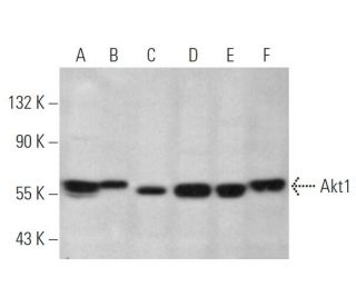
- Akt1 Anticuerpo (F-8) es un monoclonal de ratón IgG1 κ, ver las 14 publicaciones, proporcionado como 200 µg/ml
- planteada frente a un péptido que corresponde al C-terminus (h) del Akt1 de human origen
- recomendado para detectarAkt1 de mouse, rat y human origen, mediante WB, IP, IF, IHC(P) y ELISA
- Ver Akt1 (B-1): sc-5298 para Akt1 anticuerpos conjugados, incluyendo AC, HRP, FITC, PE, Alexa Fluor® 488, 594, 647, 680 y 790.
- m-IgG Fc BP-HRP y m-IgG1 BP-HRP son los reactivos de detección secundarios preferidos para Akt1 Anticuerpo (F-8) for WB and IHC(P) applications. Estos reactivos se ofrecen ahora en paquetes con Akt1 Anticuerpo (F-8)(véase la información de pedido más abajo).

ENLACES RÁPIDOS
La familia de quinasas Akt contiene varios miembros, incluyendo Akt1 (también designado como PKB o RacPK), Akt2 (también designado como PKBβ o RacPK-βb) y Akt3 (también designado como PKBγ o proto-oncogén viral de timoma 3), que muestran homología de secuencia con las familias de quinasas proteína A y C y son codificados por el proto-oncogén c-Akt. Todos los miembros de la familia Akt tienen un dominio de homología de pleckstrin. Akt1 y Akt2 son activados por la estimulación de PDGF. La activación depende de los residuos de tirosina 740 y 751 de PDGFR-β, que se unen a la subunidad del complejo de fosfatidilinositol 3-quinasas (PI 3-quinasas). La activación de Akt1 por insulina o factor de crecimiento similar a la insulina-1 (IGF-1) resulta en la fosforilación tanto de Treonina 308 como de Serina 473. La fosforilación de ambos residuos es importante para generar un alto nivel de actividad de Akt1. La fosforilación de Treonina 308 no depende de la fosforilación de Serina 473 in vivo. Por lo tanto, las proteínas Akt se fosforilan y se activan en células estimuladas por insulina/IGF-1 por una quinasa(s) aguas arriba. La activación de Akt1 y Akt2 es inhibida por el inhibidor de PI quinasa wortmannin, lo que sugiere que las proteínas señalizan aguas abajo de las quinasas PI.
Información sobre pedidos
| Nombre del producto | Número de catálogo | UNIDAD | Precio | CANTIDAD | Favoritos | |
Akt1 Anticuerpo (F-8) | sc-271149 | 200 µg/ml | $322.00 | |||
Paquete de Akt1 (F-8): m-IgG Fc BP-HRP | sc-540213 | 200 µg Ab; 10 µg BP | $361.00 | |||
Paquete de Akt1 (F-8): m-IgG1 BP-HRP | sc-541975 | 200 µg Ab; 20 µg BP | $361.00 | |||
Akt1 (F-8) péptido neutralizante | sc-271149 P | 100 µg/0.5 ml | $69.00 |