")
Anticorps NG2 (LHM 2): sc-53389
- L'anticorps NG2 LHM 2 est un monoclonal IgG1 κ L'Anticorps NG2, cité dans 23 publications, fourni en 200 µg/ml
- élevé contre l'extrait brut de cellules A375P d'origine human.
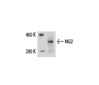
- L'Anticorps NG2 (LHM 2) est recommandé pour la détection de NG2 d'origine mouse, rat et human par WB, IP, IF, IHC(P) et FCM
- Anti-L'Anticorps NG2 (LHM 2) est disponible conjugué à l'agarose pour IP; à l'HRP pour WB, IHC(P) et ELISA; et soit à la phycoerythrin ou FITC pour IF, IHC(P) et FCM
- aussi disponible conjugué à l'Alexa Fluor® 488, Alexa Fluor® 546, Alexa Fluor® 594 ou Alexa Fluor® 647 pour WB (RGB), IF, IHC(P) et FCM
- aussi disponible conjugué à l'Alexa Fluor® 680 ou Alexa Fluor® 790 pour WB (NIR), IF et FCM
- m-IgG Fc BP-HRP et m-IgG1 BP-HRP sont les réactifs de détection secondaire préférés pour NG2 Antibody (LHM 2) for WB and IHC(P) applications. Ces réactifs sont désormais proposés en lots avec NG2 Antibody (LHM 2)(voir les informations de commande ci-dessous).

ACCÈS RAPIDE AUX LIENS
VOIR ÉGALEMENT...
L'anticorps NG2 (LHM 2) est un anticorps monoclonal IgG1 à chaîne légère kappa de souris qui détecte la protéine NG2 dans des échantillons de souris, de rats et d'humains par western blotting (WB), immunoprécipitation (IP), immunofluorescence (IF), immunohistochimie et cytométrie de flux (FCM). L'anticorps NG2 (LHM 2) se présente sous des formes non conjuguées et sous divers conjugués, notamment l'agarose, la peroxydase de raifort (HRP), la phycoérythrine (PE), l'isothiocyanate de fluorescéine (FITC) et de multiples options Alexa Fluor®. NG2, également connu sous le nom de protéoglycane 4 de chondroïtine sulfate associé au mélanome, stabilise les interactions cellule-substratum lors de l'étalement précoce des cellules de mélanome sur les membranes basales endothéliales. Cette stabilisation favorise la progression du mélanome primaire en renforçant l'activation des voies de signalisation essentielles à l'invasion et à la croissance de la tumeur. La phosphorylation de la thréonine 2256 dans la NG2 de rat (ou de la thréonine 2252 dans la NG2 humaine) déclenche la redistribution sur les surfaces des astrocytomes, la polarisation des cellules et l'augmentation de leur motilité. La NG2 fonctionne comme un corécepteur dans les cellules de mélanome malin, favorisant l'étalement et la formation de contacts focaux avec l'intégrine α4 β1. NG2 apparaît sur les vaisseaux sanguins des embryons de rats et sur les cellules endothéliales des microvaisseaux du système nerveux central, tout en servant de nouveau marqueur pour les cellules souches épidermiques, contribuant à leur distribution et à leur regroupement.
Alexa Fluor® est une marque déposée de Molecular Probes Inc., OR., USA
LI-COR® et Odyssey® sont marques déposées de LI-COR Biosciences
Anticorps NG2 (LHM 2) Références:
- Le récepteur du PDGF (alpha) ne répond pas au PDGF-AA dans les cellules musculaires lisses aortiques de la souris knock-out NG2. | Grako, KA., et al. 1999. J Cell Sci. 112 (Pt 6): 905-15. PMID: 10036240
- Isolement d'anticorps humains spécifiques de tumeurs par sélection d'une bibliothèque de phages d'anticorps sur des cellules de mélanome. | Kupsch, JM., et al. 1999. Clin Cancer Res. 5: 925-31. PMID: 10213230
- Les cellules progénitrices de l'épiderme et du follicule pileux expriment la protéine centrale du protéoglycane de sulfate de chondroïtine associé au mélanome. | Ghali, L., et al. 2004. J Invest Dermatol. 122: 433-42. PMID: 15009727
- Les cellules exprimant le NG2 dans la zone subventriculaire sont des cellules de type C et contribuent à la génération d'interneurones dans l'hippocampe postnatal. | Aguirre, AA., et al. 2004. J Cell Biol. 165: 575-89. PMID: 15159421
- Le protéoglycane NG2 favorise la motilité des cellules endothéliales et l'angiogenèse par l'engagement de la galectine-3 et de l'intégrine alpha3beta1. | Fukushi, J., et al. 2004. Mol Biol Cell. 15: 3580-90. PMID: 15181153
- La phosphorylation du protéoglycane NG2 par la protéine kinase C-alpha régule la distribution membranaire polarisée et la motilité cellulaire. | Makagiansar, IT., et al. 2004. J Biol Chem. 279: 55262-70. PMID: 15504744
- Neurogenèse postnatale et gliogenèse dans le bulbe olfactif à partir de progéniteurs exprimant NG2 dans la zone sous-ventriculaire. | Aguirre, A. and Gallo, V. 2004. J Neurosci. 24: 10530-41. PMID: 15548668
- Dysmorphogenèse des capillaires péritubulaires corticaux du rein chez les souris déficientes en angiopoiétine-2. | Pitera, JE., et al. 2004. Am J Pathol. 165: 1895-906. PMID: 15579434
- Innervation des fibres grimpantes des cellules gliales exprimant NG2 dans le cervelet des mammifères. | Lin, SC., et al. 2005. Neuron. 46: 773-85. PMID: 15924863
- Participation du protéoglycane NG2 dans les réponses des cellules musculaires lisses de l'aorte de rat au facteur de croissance dérivé des plaquettes. | Grako, KA. and Stallcup, WB. 1995. Exp Cell Res. 221: 231-40. PMID: 7589250
- Propagation et formation de contacts focaux de cellules de mélanome humain en réponse à la stimulation du protéoglycane associé au mélanome (NG2) et de l'intégrine alpha 4 bêta 1. | Iida, J., et al. 1995. Cancer Res. 55: 2177-85. PMID: 7743521
- Génération et sélection d'anticorps monoclonaux, de Fv à chaîne unique et de phages de fusion d'anticorps spécifiques d'antigènes associés au mélanome humain. | Kupsch, JM., et al. 1995. Melanoma Res. 5: 403-11. PMID: 8589614
Informations pour la commande
| Nom du produit | Ref. Catalogue | COND. | Prix HT | QTÉ | Favoris | |
Anticorps NG2 (LHM 2) | sc-53389 | 200 µg/ml | $322.00 | |||
NG2 (LHM 2): m-IgG Fc BP-HRP Kit | sc-526805 | 200 µg Ab; 10 µg BP | $361.00 | |||
NG2 (LHM 2): m-IgG1 BP-HRP Kit | sc-532178 | 200 µg Ab; 20 µg BP | $361.00 | |||
Anticorps NG2 (LHM 2) AC | sc-53389 AC | 500 µg/ml, 25% agarose | $424.00 | |||
Anticorps NG2 (LHM 2) HRP | sc-53389 HRP | 200 µg/ml | $322.00 | |||
Anticorps NG2 (LHM 2) FITC | sc-53389 FITC | 200 µg/ml | $336.00 | |||
Anticorps NG2 (LHM 2) PE | sc-53389 PE | 200 µg/ml | $349.00 | |||
Anticorps NG2 (LHM 2) Alexa Fluor® 488 | sc-53389 AF488 | 200 µg/ml | $364.00 | |||
Anticorps NG2 (LHM 2) Alexa Fluor® 546 | sc-53389 AF546 | 200 µg/ml | $364.00 | |||
Anticorps NG2 (LHM 2) Alexa Fluor® 594 | sc-53389 AF594 | 200 µg/ml | $364.00 | |||
Anticorps NG2 (LHM 2) Alexa Fluor® 647 | sc-53389 AF647 | 200 µg/ml | $364.00 | |||
Anticorps NG2 (LHM 2) Alexa Fluor® 680 | sc-53389 AF680 | 200 µg/ml | $364.00 | |||
Anticorps NG2 (LHM 2) Alexa Fluor® 790 | sc-53389 AF790 | 200 µg/ml | $364.00 |