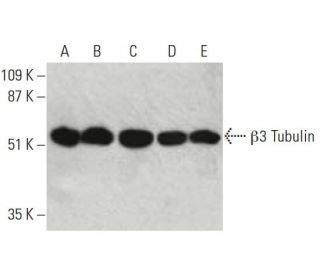
Date published: 2025-9-6

021-6093-6350

SCBT Portrait Logo
Seach Input

β3 Tubulin 抗体 (AA10): sc-80016

4.9(9)
寫評論提問

说明书
  • β3 Tubulin抗体(AA10)是小鼠单克隆IgG2a κ, 在36篇文献中引用,规格为200 µg/ml
  • 与rat的β3 Tubulin相比,具有与氨基酸436-450的表位图谱
  • 抗-beta 3 Tubulin 抗体 (AA10) 推荐用于 WB, IP 和 IF,检测mouse, rat 和human 来源的 β3 Tubulin
  • 抗beta 3 Tubulin抗体(AA10)可与琼脂糖结合用于IP;与HRP结合用于WB、IHC(P)和ELISA;与藻红蛋白FITC结合用于IF、IHC(P)和FCM
  • 还可偶联Alexa Fluor® 488, Alexa Fluor® 546, Alexa Fluor® 594Alexa Fluor® 647,用于WB (RGB), IF, IHC(P) 和 FCM, 以及用于RGB荧光成像系统,例如iBright™ FL1000, FluorChem™, Typhoon, Azure和其他类似的系统
  • 还可偶联Alexa Fluor® 680Alexa Fluor® 790, 用于WB (NIR), IF 和 FCM; 以及用于近红外(NIR)检测系统,如LI-COR®/Odyssey®, iBright™ FL1000, FluorChem™, Typhoon, Azure和类似系统
  • 关于如何获取β3 Tubulin (AA10): sc-80016免费10 µg小样,联系我们技术服务部门 (或者您当地的代理商)了解详情。
  • 2a BP-HRP">m-IgG2a BP-HRP是β3 Tubulin Antibody (AA10) 适用于 WB 应用。 的首选辅助检测试剂。该试剂现与β3 Tubulin Antibody (AA10) 打包提供(请参阅下面的订购信息)。

快捷链接

関連項目

β3 管蛋白抗体(AA10)是一种小鼠单克隆 IgG2a kappa 轻链抗体,可通过应用诸如蛋白质印迹(WB)、免疫沉淀(IP)和免疫荧光(IF)等方法检测小鼠、大鼠和人类样本中的 β3 管蛋白。抗-β3 管 抗体(AA10)有非结合型和多种结合型,包括琼脂糖、辣根过氧化物酶(HRP)、藻红蛋白(PE)、异硫氰酸荧光素(FITC)和多种Alexa Fluor®结合型。β3微管蛋白是一种神经元特异性细胞骨架蛋白,在微管的形成和稳定中起着至关重要的作用,而微管对于维持细胞形状、细胞内运输和神经元过程的正常运作至关重要。β3微管蛋白在神经元中的独特表达(与其他β微管蛋白同源物不同)凸显了其在神经发育和维持神经元完整性方面的重要性。β3微管蛋白与多种蛋白相互作用,包括MAP2和Tau,它们对于微管稳定和神经信号传导至关重要。了解β3微管蛋白的相互作用和功能对于阐明神经退行性疾病和神经元损伤的机制至关重要,因此β3微管蛋白(AA10)单克隆抗体成为研究神经系统研究人员的重要工具。

仅限研究使用。不适用于诊断和治疗用途。

Alexa Fluor® 是Molecular Probes Inc., OR., USA的商标

LI-COR®和 Odyssey® 是LI-COR Biosciences的注册商标

订购信息

产品名称产品编号规格价格数量收藏夹

β3 Tubulin 抗体 (AA10)

sc-80016
200 µg/ml
$316.00

β3 Tubulin (AA10): m-IgG2a BP-HRP 套装

sc-545917
200 µg Ab; 10 µg BP
$354.00

β3 Tubulin 抗体 (AA10) AC

sc-80016 AC
500 µg/ml, 25% agarose
$416.00

β3 Tubulin 抗体 (AA10) HRP

sc-80016 HRP
200 µg/ml
$316.00

β3 Tubulin 抗体 (AA10) FITC

sc-80016 FITC
200 µg/ml
$330.00

β3 Tubulin 抗体 (AA10) PE

sc-80016 PE
200 µg/ml
$343.00

β3 Tubulin 抗体 (AA10) Alexa Fluor® 488

sc-80016 AF488
200 µg/ml
$357.00

β3 Tubulin 抗体 (AA10) Alexa Fluor® 546

sc-80016 AF546
200 µg/ml
$357.00

β3 Tubulin 抗体 (AA10) Alexa Fluor® 594

sc-80016 AF594
200 µg/ml
$357.00

β3 Tubulin 抗体 (AA10) Alexa Fluor® 647

sc-80016 AF647
200 µg/ml
$357.00

β3 Tubulin 抗体 (AA10) Alexa Fluor® 680

sc-80016 AF680
200 µg/ml
$357.00

β3 Tubulin 抗体 (AA10) Alexa Fluor® 790

sc-80016 AF790
200 µg/ml
$357.00

I need to use an antibody to detect the protein in samples of porcine origin. Do you know if this mouse monoclonal antibody will work?

Asked by: jerojero
Please contact our Technical Service Department and we would be happy to perform sequence analysis and review our data to answer any questions you may have about additional species reactivity.
Answered by: Tech Service 3
Date published: 2017-02-01
  • y_2025, m_8, d_27, h_5CST
  • bvseo_bulk, prod_bvqa, vn_bulk_3.0.42
  • cp_1, bvpage1
  • co_hasquestionsanswers, tq_1
  • loc_zh_CN, sid_80016, prod, sort_[SortEntry(order=LAST_APPROVED_ANSWER_SUBMISSION_TIME, direction=DESCENDING)]
  • clientName_scbt
  • bvseo_sdk, java_sdk, bvseo-4.0.0
  • CLOUD, getContent, 105ms
  • QUESTIONS, PRODUCT
Rated 5 out of 5 by from Great signalStrong signal, no background, band at 55 KDa. Tested in mouse hippocampal lysates and rat primary cultured neurons (1:500 in 5% nonfat dry milk-TBS-Tween).
Date published: 2023-04-07
Rated 5 out of 5 by from Great signalStrong signal, no background, band at 55 KDa. Tested in mouse hippocampal lysates and rat primary cultured neurons (1:500 in 5% nonfat dry milk-TBS-Tween).
Date published: 2023-04-04
Rated 5 out of 5 by from Great signalStrong signal, no background, band at 55 KDa. Tested in mouse hippocampal lysates and rat primary cultured neurons (1:500 in 5% nonfat dry milk-TBS-Tween).
Date published: 2023-04-04
Rated 5 out of 5 by from Great signalStrong signal, no background, band at 55 KDa. Tested in mouse hippocampal lysates and rat primary cultured neurons (1:500 in 5% nonfat dry milk-TBS-Tween).
Date published: 2023-04-04
Rated 5 out of 5 by from Excelent antibodyStrong signal, no background, band at 55 KDa. Tested in mouse hippocampal lysates and rat primary cultured neurons (1:500 in 5% nonfat dry milk-TBS-Tween).
Date published: 2023-04-04
Rated 4 out of 5 by from Melanocortin 4 receptor activation protects striatα-Melanocyte stimulating hormone (α-MSH) is a melanocortin which exerts potent anti-inflammatory and antiapoptotic effects. Melanocortin 4 receptors (MC4R) are abundantly expressed in the brain and we previously demonstrated that [Nle(4), D-Phe(7)]melanocyte-stimulating hormone (NDP-MSH), an α-MSH analogue, increased expression of brain derived-neurotrophic factor (BDNF), and peroxisome proliferator-activated receptor- γ (PPAR-γ). We hypothesized that melanocortins could affect striatal cell survival through BDNF and PPAR-γ. First, we determined the expression of these factors in the striatum. Acute intraperitoneal administration (0.5 mg/kg) of α-MSH increased the levels of BDNF mRNA in rat striatum but not in rat cerebral cortex. Also, protein expression of PPAR-γ and MC4R was increased by acute treatment with α-MSH in striatum but not in cortex. No changes were observed by 48 h treatment. Next, we evaluated melanocortins effect on neuron and glial survival. 3-nitropropionic acid (3-NP), which is known to induce striatal degeneration, was used to induce cell death in the rat striatal cell line ST14A expressing mutant human huntingtin (Q120) or in ST14A cells expressing normal human huntingtin (Q15), in primary cultured astrocytes, and in BV2 cells. NDP-MSH protected Q15 cells, astrocytes and BV2 cells from death by 3-NP whereas it did not fully protect Q120 cells. Protection of Q15 cells and astrocytes was blocked by a MC4R specific inhibitor (JKC-363) and a PPAR-γ antagonist (GW9662). The BDNF receptor antagonist (ANA-12) abolished NDP-MSH protective effect in astrocytes but not in Q15 cells. We demonstrate for the first time that melanocortins, acting through PPAR-γ and BDNF, protect neurons and glial cells from 3-NP toxicity
Date published: 2018-12-13
Rated 5 out of 5 by from Very good resultbeta-3-tubulin antibody (AA10) sc-800166: Immunofluorescence staining of beta-3-tubulin (green) and S100 (red) in rat dorsal root ganglion fixed in 4% paraformaldehyde.
Date published: 2018-02-06
Rated 5 out of 5 by from Produced good western blot bandThe tubulin antibody produced a clear western blot band for our cell lysate
Date published: 2016-12-20
  • y_2025, m_8, d_27, h_5
  • bvseo_bulk, prod_bvrr, vn_bulk_3.0.42
  • cp_1, bvpage1
  • co_hasreviews, tv_0, tr_9
  • loc_zh_CN, sid_80016, prod, sort_[SortEntry(order=SUBMISSION_TIME, direction=DESCENDING)]
  • clientName_scbt
  • bvseo_sdk, java_sdk, bvseo-4.0.0
  • CLOUD, getReviews, 19ms
  • REVIEWS, PRODUCT
β3 Tubulin 抗体 (AA10) is rated 4.9 out of 5 by 9.
  • y_2025, m_8, d_27, h_5
  • bvseo_bulk, prod_bvrr, vn_bulk_3.0.42
  • cp_1, bvpage1
  • co_hasreviews, tv_0, tr_9
  • loc_zh_CN, sid_80016, prod, sort_[SortEntry(order=SUBMISSION_TIME, direction=DESCENDING)]
  • clientName_scbt
  • bvseo_sdk, java_sdk, bvseo-4.0.0
  • CLOUD, getAggregateRating, 107ms
  • REVIEWS, PRODUCT