")
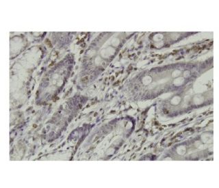
: sc-100762. Immunoperoxidase staining of formalin-fixed, paraffin-embedded human small Intestine tissue showing nuclear and cytoplasmic localization.")
: sc-100762. Western blot analysis of RGS3 expression in non-transfected: sc-117752 (A) and human RGS3 transfected: sc-115637 (B) 293T whole cell lysates.")
RGS3 Antibody (CC-Q7): sc-100762
- RGS3 Antibody (CC-Q7) is a mouse monoclonal IgG1 κ RGS3 antibody, cited in 1 publications, provided at 100 µg/ml
- raised against recombinant RGS3 of human origin
- recommended for detection of RGS3 of human origin by WB, IP, IF, IHC(P) and ELISA
- m-IgG Fc BP-HRP and m-IgG1 BP-HRP are the preferred secondary detection reagents for RGS3 Antibody (CC-Q7) for WB and IHC(P) applications. These reagents are now offered in bundles with RGS3 Antibody (CC-Q7) (see ordering information below).

RGS3 Antibody (CC-Q7) is a mouse monoclonal IgG1 kappa light chain antibody that detects RGS3 protein of human origin by western blotting (WB), immunoprecipitation (IP), immunofluorescence (IF), immunohistochemistry, and enzyme-linked immunosorbent assay (ELISA). Anti-RGS3 antibody (CC-Q7) is available as the non-conjugated form. RGS3 plays a crucial role in the regulation of G protein signaling pathways, which are essential for transmitting signals from cell surface receptors to various intracellular effectors. Specifically, RGS3 interacts with the activated form of Gα11, a member of the heterotrimeric G protein family. This interaction enhances the GTPase activity of Gα11, leading to its inactivation and a subsequent decrease in intracellular calcium levels, which is vital for numerous cellular processes, including muscle contraction and neurotransmitter release. RGS3 is predominantly expressed in adult kidney and myocardium, where RGS3 is primarily localized in the cytoplasm. Upon activation of Gα11, RGS3 translocates to the plasma membrane, positioning RGS3 strategically to modulate G protein signaling effectively. This dynamic localization and interaction underscore RGS3′s importance in maintaining cellular homeostasis and responding to extracellular signals.
Alexa Fluor® is a trademark of Molecular Probes Inc., OR., USA
LI-COR® and Odyssey® are registered trademarks of LI-COR Biosciences
RGS3 Antibody (CC-Q7) References:
- Modulation of renal tubular cell function by RGS3. | Grüning, W., et al. 1999. Am J Physiol. 276: F535-43. PMID: 10198412
- A G-protein signaling network mediated by an RGS protein. | Guan, KL. and Han, M. 1999. Genes Dev. 13: 1763-7. PMID: 10421628
- Structural elements of G alpha subunits that interact with G beta gamma, receptors, and effectors. | Conklin, BR. and Bourne, HR. 1993. Cell. 73: 631-41. PMID: 8388779
- Inhibition of G-protein-mediated MAP kinase activation by a new mammalian gene family. | Druey, KM., et al. 1996. Nature. 379: 742-6. PMID: 8602223
- A truncated form of RGS3 negatively regulates G protein-coupled receptor stimulation of adenylyl cyclase and phosphoinositide phospholipase C. | Chatterjee, TK., et al. 1997. J Biol Chem. 272: 15481-7. PMID: 9182581
- Genomic organization, 5'-flanking region, and chromosomal localization of the human RGS3 gene. | Chatterjee, TK., et al. 1997. Genomics. 45: 429-33. PMID: 9344672
- RGS3 inhibits G protein-mediated signaling via translocation to the membrane and binding to Galpha11. | Dulin, NO., et al. 1999. Mol Cell Biol. 19: 714-23. PMID: 9858594
Ordering Information
| Product Name | Catalog # | UNIT | Price | Qty | FAVORITES | |
RGS3 Antibody (CC-Q7) | sc-100762 | 100 µg/ml | $339.00 | |||
RGS3 Antibody (CC-Q7): m-IgG Fc BP-HRP Bundle | sc-539709 | 100 µg Ab; 10 µg BP | $361.00 | |||
RGS3 Antibody (CC-Q7): m-IgG1 BP-HRP Bundle | sc-541664 | 100 µg Ab; 20 µg BP | $361.00 |