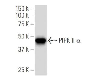
Date published: 2026-4-15

1-800-457-3801

SCBT Portrait Logo
Seach Input

PIPK II α Antibody (149.1): sc-100406

5.0(5)
Write a reviewAsk a question

Datasheets
  • PIPK II α Antibody (149.1) is a mouse monoclonal IgG1 κ PIPK II α antibody, cited in 4 publications, provided at 100 µg/ml
  • raised against recombinant PIPK II α of human origin
  • recommended for detection of PIPK II α of mouse, rat and human origin by WB, IP, IF, IHC(P) and ELISA; may cross-react with PIPK II β
  • At present, we have not yet completed the identification of the preferred secondary detection reagent(s) for PIPK II α Antibody (149.1). This work is in progress.
Gene Editing Promo Banner

QUICK LINKS

SEE ALSO...

PIPK II α Antibody (149.1) is a mouse monoclonal IgG1 kappa light chain antibody that detects PIPK II alpha protein of mouse, rat, and human origin by western blotting (WB), immunoprecipitation (IP), immunofluorescence (IF), immunohistochemistry with paraffin-embedded sections (IHCP), and enzyme-linked immunosorbent assay (ELISA). Anti-PIPK II alpha antibody (149.1) is available as the non-conjugated format. PIPK II alpha protein plays a crucial role in synthesizing phosphatidylinositol-4,5-bisphosphate, a lipid vital for various cellular processes, including cell proliferation, survival, membrane trafficking, and cytoskeletal organization. PIPK II alpha protein is predominantly located in the brain, contributing to the regulation of signaling pathways essential for neuronal function and communication. PIPK family is classified into three types, with type II PIPKs, including PIPK II α and β, being particularly important for their high expression levels in neural tissues. PIPK II alpha presence in the brain underscores significance in neurobiology, involving modulation of synaptic activity and plasticity, fundamental for learning and memory. Through facilitating phosphatidylinositol-4,5-bisphosphate production, PIPK II alpha serves as a key messenger in signal transduction pathways, making PIPK II alpha an important target for research in neurodegenerative diseases and other conditions affecting the nervous system.

For Research Use Only. Not Intended for Diagnostic or Therapeutic Use.

Alexa Fluor® is a trademark of Molecular Probes Inc., OR., USA

LI-COR® and Odyssey® are registered trademarks of LI-COR Biosciences

PIPK II α Antibody (149.1) References:

  1. The phosphatidylinositol (PI)-5-phosphate 4-kinase type II enzyme controls insulin signaling by regulating PI-3,4,5-trisphosphate degradation.  |  Carricaburu, V., et al. 2003. Proc Natl Acad Sci U S A. 100: 9867-72. PMID: 12897244
  2. Cloning and subcellular localization of a human phosphatidylinositol 3-phosphate 5-kinase, PIKfyve/Fab1.  |  Cabezas, A., et al. 2006. Gene. 371: 34-41. PMID: 16448788
  3. The mammalian phosphatidylinositol 3-phosphate 5-kinase (PIKfyve) regulates endosome-to-TGN retrograde transport.  |  Rutherford, AC., et al. 2006. J Cell Sci. 119: 3944-57. PMID: 16954148
  4. Type II PtdInsP kinases: location, regulation and function.  |  Clarke, JH., et al. 2007. Biochem Soc Symp. 149-59. PMID: 17233588
  5. Enzymes phosphorylating lipids and polysaccharides.  |  Karataeva, NA. and Nevinsky, GA. 2007. Biochemistry (Mosc). 72: 367-79. PMID: 17511601
  6. The phosphoinositide kinase PIP5K that produces the versatile signaling phospholipid PI4,5P(2).  |  Kanaho, Y., et al. 2007. Biol Pharm Bull. 30: 1605-9. PMID: 17827707
  7. Phosphatidylinositol 4-phosphate 5-kinase reduces cell surface expression of the epithelial sodium channel (ENaC) in cultured collecting duct cells.  |  Weixel, KM., et al. 2007. J Biol Chem. 282: 36534-42. PMID: 17940289
  8. Type II phosphatidylinositol 4-kinase(s) in cell signaling cascades.  |  Sinha, RK. and Subrahmanyam, G. 2007. Indian J Biochem Biophys. 44: 289-94. PMID: 18341203
  9. Beta-arrestin scaffolding of phosphatidylinositol 4-phosphate 5-kinase Ialpha promotes agonist-stimulated sequestration of the beta2-adrenergic receptor.  |  Nelson, CD., et al. 2008. J Biol Chem. 283: 21093-101. PMID: 18534983
  10. PIP4K2A regulates intracellular cholesterol transport through modulating PI(4,5)P2 homeostasis.  |  Hu, A., et al. 2018. J Lipid Res. 59: 507-514. PMID: 29353240
  11. PIP5K1C phosphoinositide kinase deficiency distinguishes PIKFYVE-dependent cancer cells from non-malignant cells.  |  Roy, A., et al. 2023. Autophagy. 19: 2464-2484. PMID: 36803256
  12. Exploring the Potential Role of Oligodendrocyte-Associated PIP4K2A in Alzheimer's Disease Complicated with Type 2 Diabetes Mellitus via Multi-Omic Analysis.  |  Nguyen, DPQ., et al. 2024. Int J Mol Sci. 25: PMID: 38928345

Ordering Information

Product NameCatalog #UNITPriceQtyFAVORITES

PIPK II α Antibody (149.1)

sc-100406
100 µg/ml
$339.00

Can sc-100406: PIPK II α (149.1) monoclonal antibody be used to stain formalin-fixed, paraffin-embedded (FFPE) tissue sections?

Asked by: jerojero
Thank you for your question. Yes, sc-100406: PIPK II α (149.1) is recommended for use in IHC with paraffin-embedded sections. We recommend performing antigen retrieval with sodium citrate buffer (pH 6) and heat. The full protocol can be found here: https://www.scbt.com/scbt/resources/protocols/immunoperoxidase-staining
Answered by: Technical Support
Date published: 2017-03-24
  • y_2026, m_4, d_9, h_7CST
  • bvseo_bulk, prod_bvqa, vn_bulk_3.0.43
  • cp_1, bvpage1
  • co_hasquestionsanswers, tq_1
  • loc_en_US, sid_100406, prod, sort_[SortEntry(order=LAST_APPROVED_ANSWER_SUBMISSION_TIME, direction=DESCENDING)]
  • clientName_scbt
  • bvseo_sdk, java_sdk, bvseo-4.0.0
  • CLOUD, getContent, 95ms
  • QUESTIONS, PRODUCT
Rated 5 out of 5 by from Great dataThis antibody will provide you with the desired results in staining and blotting. Staining is clear and specific and blotting has strong bands at the expected weight. This antibody has been used to publish.
Date published: 2017-02-08
Rated 5 out of 5 by from Performs well in WesternPerforms well in Western, good signal with mouse PIPK II transfected 293T whole cell lysate. -SCBT QC
Date published: 2015-05-25
Rated 5 out of 5 by from Exceptional immunoperoxidase nuclear stainingExceptional immunoperoxidase nuclear staining in formalin-fixed, paraffin-embedded human small Intestine tissue. -SCBT QC
Date published: 2015-03-26
Rated 5 out of 5 by from Great antibody for WBGreat antibody for WB, works in K-562 whole cell lysate. -SCBT QC
Date published: 2015-03-16
Rated 5 out of 5 by from Band detected at expected mw in HeLa wholeBand detected at expected mw in HeLa whole cell lysate. -SCBT QC
Date published: 2015-03-04
  • y_2026, m_4, d_9, h_7
  • bvseo_bulk, prod_bvrr, vn_bulk_3.0.43
  • cp_1, bvpage1
  • co_hasreviews, tv_0, tr_5
  • loc_en_US, sid_100406, prod, sort_[SortEntry(order=SUBMISSION_TIME, direction=DESCENDING)]
  • clientName_scbt
  • bvseo_sdk, java_sdk, bvseo-4.0.0
  • CLOUD, getReviews, 20ms
  • REVIEWS, PRODUCT
PIPK II α Antibody (149.1) is rated 5.0 out of 5 by 5.
  • y_2026, m_4, d_9, h_7
  • bvseo_bulk, prod_bvrr, vn_bulk_3.0.43
  • cp_1, bvpage1
  • co_hasreviews, tv_0, tr_5
  • loc_en_US, sid_100406, prod, sort_[SortEntry(order=SUBMISSION_TIME, direction=DESCENDING)]
  • clientName_scbt
  • bvseo_sdk, java_sdk, bvseo-4.0.0
  • CLOUD, getAggregateRating, 124ms
  • REVIEWS, PRODUCT