")
Anticorps pan CEA (H-8): sc-48364
- L'anticorps pan CEA H-8 est un monoclonal IgG1 κ L'Anticorps pan CEA, cité dans 4 publications, fourni en 200 µg/ml
- dirigé contre les acides aminés 35-334 situés près du N-terminus de CEA (carcinoembryonic antigen) de human
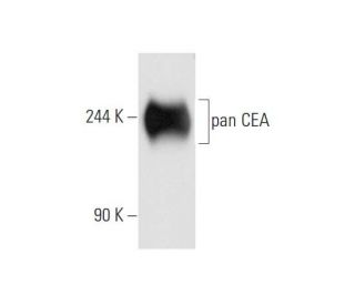
- L'Anticorps pan CEA (H-8) est recommandé pour la détection de pan CEA d'origine human par WB, IP, IF, IHC(P) et ELISA
- Anti-L'Anticorps pan CEA (H-8) est disponible conjugué à l'agarose pour IP; à l'HRP pour WB, IHC(P) et ELISA; et soit à la phycoerythrin ou FITC pour IF, IHC(P) et FCM
- aussi disponible conjugué à l'Alexa Fluor® 488, Alexa Fluor® 546, Alexa Fluor® 594 ou Alexa Fluor® 647 pour WB (RGB), IF, IHC(P) et FCM
- aussi disponible conjugué à l'Alexa Fluor® 680 ou Alexa Fluor® 790 pour WB (NIR), IF et FCM
- Nous testons actuellement encore nos anticorps secondaires pour trouver la meilleure protéine de liaison pour cet anticorps primaire pan CEA (H-8). Veuillez nous contacter si vous avez des questions concernant les réactifs de détection secondaire appropriés.

ACCÈS RAPIDE AUX LIENS
VOIR ÉGALEMENT...
L'anticorps anti-pan CEA (H-8) est un anticorps monoclonal IgG1 à chaîne légère kappa de souris qui détecte la protéine pan CEA d'origine humaine par western blotting (WB), immunoprécipitation (IP), immunofluorescence (IF), immunohistochimie sur coupes extraites de paraffine (IHCP) et enzyme-linked immunosorbent assay (ELISA). L'anticorps anti-pan CEA (H-8) est disponible sous forme non conjuguée et sous diverses formes conjuguées, notamment agarose, peroxydase de raifort (HRP), phycoérythrine (PE), isothiocyanate de fluorescéine (FITC) et plusieurs conjugués Alexa Fluor®. L'antigène carcino-embryonnaire (CEA) est un marqueur tumoral essentiel largement utilisé dans les immunodosages sériques pour la détection des carcinomes, jouant un rôle important dans le diagnostic et le suivi du cancer. L'anticorps pan CEA (H-8) cible les protéines principalement situées à la surface des cellules, où le CEA est impliqué dans les processus d'adhésion cellulaire essentiels au maintien de l'architecture des tissus et à la facilitation de la communication entre les cellules. Cette localisation est particulièrement importante en biologie tumorale, car les altérations de l'adhésion cellulaire peuvent entraîner une augmentation de la capacité d'invasion des tumeurs et des métastases. Les protéines CEACAM, y compris le pan CEA, sont codées par des gènes regroupés sur le bras long du chromosome 19, et les niveaux d'expression peuvent fournir des indications précieuses sur la progression de diverses tumeurs malignes.
Alexa Fluor® est une marque déposée de Molecular Probes Inc., OR., USA
LI-COR® et Odyssey® sont marques déposées de LI-COR Biosciences
Anticorps pan CEA (H-8) Références:
- Augmentation de l'expression du transgène à partir du promoteur de l'antigène carcinoembryonnaire (CEA) via un système de régulation du gène GAL4. | Koch, PE., et al. 2001. Mol Ther. 3: 278-83. PMID: 11273768
- Kératine et antigène carcinoembryonnaire (CEA) dans les cellules de mélanome humain. | Om, A., et al. 1991. Virchows Arch B Cell Pathol Incl Mol Pathol. 61: 81-7. PMID: 1720588
- Diversité haplotypique des gènes CEACAM humains: effets sur la susceptibilité à la maladie méningococcique. | Callaghan, MJ., et al. 2008. Genes Immun. 9: 30-7. PMID: 17960155
- Le domaine transmembranaire de CEACAM1, mais pas le domaine cytoplasmique, dirige l'internalisation des pathogènes humains via des microdomaines membranaires. | Muenzner, P., et al. 2008. Cell Microbiol. 10: 1074-92. PMID: 18081725
- La voie de l'apoptose médiée par le CEACAM1 est activée par le CEA et déclenche un double clivage du CEACAM1. | Nittka, S., et al. 2008. Oncogene. 27: 3721-8. PMID: 18278069
- Dynamique de CEACAM1 pendant la suppression de l'activation des lymphocytes T CD4+ par Neisseria gonorrhoeae. | Lee, HS., et al. 2008. J Immunol. 180: 6827-35. PMID: 18453603
- Modification de l'épissage de CEACAM1 dans le cancer du sein: identification des séquences régulatrices qui contrôlent l'épissage de CEACAM1 en isoformes à domaine cytoplasmique long ou court. | Gaur, S., et al. 2008. Mol Cancer. 7: 46. PMID: 18507857
- CEACAM1, une cible transcriptionnelle directe de SOX9 identifiée dans l'épithélium du côlon. | Zalzali, H., et al. 2008. Oncogene. 27: 7131-8. PMID: 18794798
- CEACAM1 inhibe les réponses antibactériennes déclenchées par le récepteur Toll-like 2 des cellules épithéliales pulmonaires humaines. | Slevogt, H., et al. 2008. Nat Immunol. 9: 1270-8. PMID: 18836450
- L'expression de CEACAM1 induite par les cytokines sur les kératinocytes est caractéristique de la peau psoriasique et contribue à prolonger la durée de vie des neutrophiles. | Rahmoun, M., et al. 2009. J Invest Dermatol. 129: 671-81. PMID: 18843289
- L'interdépendance des CEACAM-1, -3, -6 et -8 induit l'adhésion des neutrophiles humains aux cellules endothéliales. | Skubitz, KM. and Skubitz, AP. 2008. J Transl Med. 6: 78. PMID: 19077207
- Démonstration tissulaire de l'antigène carcinoembryonnaire (ACE) dans la colite ulcéreuse. | Isaacson, P. 1976. Gut. 17: 561-7. PMID: 964689
Informations pour la commande
| Nom du produit | Ref. Catalogue | COND. | Prix HT | QTÉ | Favoris | |
Anticorps pan CEA (H-8) | sc-48364 | 200 µg/ml | $322.00 | |||
Anticorps pan CEA (H-8) AC | sc-48364 AC | 500 µg/ml, 25% agarose | $424.00 | |||
Anticorps pan CEA (H-8) HRP | sc-48364 HRP | 200 µg/ml | $322.00 | |||
Anticorps pan CEA (H-8) FITC | sc-48364 FITC | 200 µg/ml | $336.00 | |||
Anticorps pan CEA (H-8) PE | sc-48364 PE | 200 µg/ml | $349.00 | |||
Anticorps pan CEA (H-8) Alexa Fluor® 488 | sc-48364 AF488 | 200 µg/ml | $364.00 | |||
Anticorps pan CEA (H-8) Alexa Fluor® 546 | sc-48364 AF546 | 200 µg/ml | $364.00 | |||
Anticorps pan CEA (H-8) Alexa Fluor® 594 | sc-48364 AF594 | 200 µg/ml | $364.00 | |||
Anticorps pan CEA (H-8) Alexa Fluor® 647 | sc-48364 AF647 | 200 µg/ml | $364.00 | |||
Anticorps pan CEA (H-8) Alexa Fluor® 680 | sc-48364 AF680 | 200 µg/ml | $364.00 | |||
Anticorps pan CEA (H-8) Alexa Fluor® 790 | sc-48364 AF790 | 200 µg/ml | $364.00 |