")
: sc-71569. C32 全细胞裂解液中 Melan-A 表达的 Western 印迹分析.")
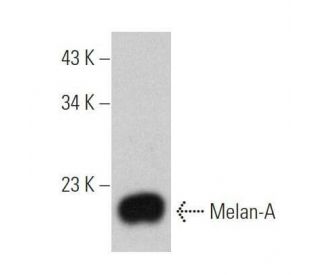
: sc-71569. Western 印迹分析 SK-MEL-28 全细胞裂解液中 Melan-A 的表达.")
Melan-A 抗体 (0. N. 397): sc-71569. C32 全细胞裂解液中 Melan-A 表达的 Western 印迹分析.
Melan-A 抗体 (0.N.397): sc-71569
- Melan-A 抗体 0.N.397 是小鼠单克隆 IgG2b,提供 200 µg/ml
- 免疫human物种的重组Melan-A
- 推荐用于 mouse, rat 和 human 来源的Melan-A WB, IP, IF 和 IHC(P)检测
- 看 Melan-A (A103): sc-20032,可查看有标记的 Melan-A 抗体,包括AC, HRP, FITC, PE, Alexa Fluor® 488, 594, 647, 680 和 790
- 目前,我们还没有完成Melan-A 抗体 (0.N.397)的首选二抗检测试剂的鉴定。这项工作正在进行中。

快捷链接
相关产品
描述
基因信息
说明书与实验方案
研究信息
関連項目
细胞毒性T淋巴细胞(CTL)识别的黑色素瘤相关抗原分为三类:黑色素细胞分化抗原、癌症/测试特异性抗原和突变或异常表达抗原。许多这些抗原由HLA分子呈递给T细胞的肽组成;它们代表了癌症免疫疗法的潜在靶点。Melan-A(也称为MART-1)是一种黑色素细胞分化抗原,对黑色素瘤、黑色素细胞系和视网膜具有特异性。黑色素A肽被黑色素瘤患者中大多数HLA-A2限制性肿瘤特异性肿瘤浸润淋巴细胞识别。抗黑色素瘤细胞毒性T淋巴细胞可以由Melan-A肽产生,表明Melan-A是黑色素瘤患者抗原特异性免疫治疗的潜在候选者。
仅限研究使用。不适用于诊断和治疗用途。
Alexa Fluor® 是Molecular Probes Inc., OR., USA的商标
LI-COR®和 Odyssey® 是LI-COR Biosciences的注册商标
Melan-A 抗体 (0.N.397) 参考文献:
- HMB45阴性黑色素瘤中MITF、酪氨酸酶、黑色素-A和MAGE-1的免疫图谱。 | Xu, X., et al. 2002. Am J Surg Pathol. 26: 82-7. PMID: 11756773
- 肽特异性 CD8+ T 细胞在体内的进化:对 Melan-A/MART-1 肽疫苗的反应。 | Jäger, E., et al. 2002. Int J Cancer. 98: 376-88. PMID: 11920589
- 封装到立体稳定脂质体中可增强黑色素瘤相关 Melan-A/MART-1 表位的免疫原性。 | Adamina, M., et al. 2004. Br J Cancer. 90: 263-9. PMID: 14710238
- 对 Melan-a/MART-1 肿瘤蛋白抗原性的多肽计算分析。 | Tiwari, R., et al. 2004. Peptides. 25: 1865-71. PMID: 15501517
- 黑色素瘤相关抗原酪氨酸酶而非Melan-A/MART-1的表达和呈递在热休克反应期间发生分离。 | Milani, V., et al. 2005. Int Immunol. 17: 257-68. PMID: 15642953
- 建立可上调黑色素瘤抗原 Melan-A/MART-1 的化学品筛选系统。 | Song, HZ., et al. 2009. Tohoku J Exp Med. 217: 231-7. PMID: 19282659
- CD4+T淋巴细胞识别的HLA-DPB1*0501限制性Melan-A/MART-1表位的鉴定:在亚洲人群中的免疫疗法应用前景。 | Meng, Z., et al. 2011. J Immunother. 34: 525-34. PMID: 21760531
- 循环 CD4+ T 细胞在黑色素-A 的刺激下产生 IL4 或 IL17,而在 NY-ESO-1 的刺激下不产生 IL4 或 IL17,这对 IV 期黑色素瘤患者的生存有负面影响。 | Zelba, H., et al. 2014. Clin Cancer Res. 20: 4390-9. PMID: 24938524
- 犬髓外浆细胞瘤中的 Melan-A 免疫标记。 | Schuwerk, L., et al. 2024. Vet Pathol. 61: 904-911. PMID: 38642035
- 肾上腺皮质肿瘤和其他类固醇肿瘤对黑色素-A(Mart-1)抗体 A103 的免疫反应。 | Busam, KJ., et al. 1998. Am J Surg Pathol. 22: 57-63. PMID: 9422316
订购信息
| 产品名称 | 产品编号 | 规格 | 价格 | 数量 | 收藏夹 | |
Melan-A 抗体 (0.N.397) | sc-71569 | 200 µg/ml | $322.00 |