")
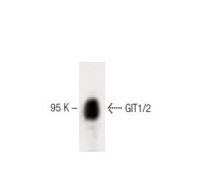
: sc-135925. Western blot analysis of GIT1/2 expression in rat cerebrum tissue extract.")
: sc-135925. Immunofluorescence staining of NIH/3T3 cells showing cytoplasmic localization.")
: sc-135925. Western blot analysis of GIT1/2 expression in untreated HeLa (A), chemically-treated HeLa (B), untreated HCT-116 (C) and chemically-treated HCT-116 (D, E) whole cell lysates. β-Actin (C4): sc-47778 used as loading control. Detection reagent used: m-IgG Fc BP-HRP: sc-525409.")
GIT1/2 Antibody (13): sc-135925
- GIT1/2 Antibody (13) is a mouse monoclonal IgG1 GIT1/2 antibody, cited in 2 publications, provided at 50 µg/0.5 ml
- raised against amino acids 140-472 of GIT2 of chicken origin
- recommended for detection of GIT1 and GIT2 of mouse, rat, human and avian origin by WB, IP and IF
- m-IgG Fc BP-HRP is the preferred secondary detection reagent for GIT1/2 Antibody (13) for WB applications. This reagent is now offered in a bundle with GIT1/2 Antibody (13) (see ordering information below).

GIT1/2 Antibody (13) is a mouse monoclonal IgG1 antibody that detects GIT1/2 protein of mouse, rat, human, and avian origin by western blotting (WB), immunoprecipitation (IP), and immunofluorescence (IF). Anti-GIT1/2 antibody (13) is available as the non-conjugated format. GIT1 and GIT2 proteins play a crucial role in cellular signaling and vesicular trafficking, acting as GTPase-activating proteins (GAP) for the ADP ribosylation factor (ARF) family of small GTP-binding proteins. GIT1 and GIT2 proteins are primarily located in the cytoplasm and at the plasma membrane, where GIT1 and GIT2 facilitate regulation of endocytosis and exocytosis processes. Proper localization of GIT1 and GIT2 is essential for maintaining cellular homeostasis and ensuring efficient communication between cells. Dysregulation of GIT1 and GIT2 can lead to impaired receptor signaling and has been implicated in various diseases, including cancer and metabolic disorders. Using GIT1/2 (13) monoclonal antibody in research can provide valuable insights into G protein-coupled receptor signaling dynamics and vesicular transport mechanisms, ultimately contributing to a better understanding of cellular function and disease pathology.
Alexa Fluor® is a trademark of Molecular Probes Inc., OR., USA
LI-COR® and Odyssey® are registered trademarks of LI-COR Biosciences
GIT1/2 Antibody (13) References:
- The GIT family of ADP-ribosylation factor GTPase-activating proteins. Functional diversity of GIT2 through alternative splicing. | Premont, RT., et al. 2000. J Biol Chem. 275: 22373-80. PMID: 10896954
- Characterization of the endogenous GIT1-betaPIX complex, and identification of its association to membranes. | Botrugno, OA., et al. 2006. Eur J Cell Biol. 85: 35-46. PMID: 16373173
- The multifunctional GIT family of proteins. | Hoefen, RJ. and Berk, BC. 2006. J Cell Sci. 119: 1469-75. PMID: 16598076
- GIT2 represses Crk- and Rac1-regulated cell spreading and Cdc42-mediated focal adhesion turnover. | Frank, SR., et al. 2006. EMBO J. 25: 1848-59. PMID: 16628223
- Differential expression of the ARF GAP genes GIT1 and GIT2 in mouse tissues. | Schmalzigaug, R., et al. 2007. J Histochem Cytochem. 55: 1039-48. PMID: 17565117
- The GIT-PIX complexes regulate the chemotactic response of rat basophilic leukaemia cells. | Gavina, M., et al. 2010. Biol Cell. 102: 231-44. PMID: 19912111
- Impaired spine formation and learning in GPCR kinase 2 interacting protein-1 (GIT1) knockout mice. | Menon, P., et al. 2010. Brain Res. 1317: 218-26. PMID: 20043896
- RhoJ interacts with the GIT-PIX complex and regulates focal adhesion disassembly. | Wilson, E., et al. 2014. J Cell Sci. 127: 3039-51. PMID: 24928894
- Expanding functions of GIT Arf GTPase-activating proteins, PIX Rho guanine nucleotide exchange factors and GIT-PIX complexes. | Zhou, W., et al. 2016. J Cell Sci. 129: 1963-74. PMID: 27182061
- GIT2-A keystone in ageing and age-related disease. | van Gastel, J., et al. 2018. Ageing Res Rev. 43: 46-63. PMID: 29452267
Ordering Information
| Product Name | Catalog # | UNIT | Price | Qty | FAVORITES | |
GIT1/2 Antibody (13) | sc-135925 | 50 µg/0.5 ml | $322.00 | |||
GIT1/2 Antibody (13): m-IgG Fc BP-HRP Bundle | sc-551914 | 50 µg Ab; 10 µg BP | $361.00 |