")
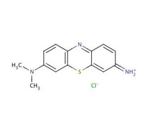

Giemsa Stain 의 분자 구조, CAS 번호: 51811-82-6
Giemsa Stain (CAS 51811-82-6)
인용논문 보기 (3)
대체 이름:
Giemsa solution
적용:
Giemsa Stain 염색질과 핵막을 위한 고품질 염색약입니다.
CAS 등록번호:
51811-82-6
분자량:
291.8
분자식:
C14H14ClN3S
연구용으로만 사용가능합니다. 진단이나 치료용으로 사용불가합니다.
* 참조분석증명서대량의 측정 데이터(함수량포함).
빠른 링크
주문정보
논문정보
설명
기술정보
안전정보
SDS 및 분석 증명서
CAS 번호 51811-82-6으로 식별되는 지엠사 염색은 염색체의 시각화 및 분화를 용이하게 하기 위해 현미경 및 세포 생물학 분야에서 널리 사용되는 고전적인 세포 유전학 기법으로, 염색체를 염색하는 데 사용됩니다. 주로 메틸렌 블루, 에오신, 하늘색 B로 구성된 이 염색은 DNA의 인산염 그룹에 선택적으로 결합하여 다른 염색체 구조와 세포 유형을 구별하는 데 중요한 뚜렷한 착색 패턴을 만들어냅니다. 염색 과정은 일반적으로 준비된 세포 슬라이드에 염색약을 도포하여 세포 내의 DNA와 RNA에 달라붙어 염색질, 핵 구조 및 세포질과 같은 특징을 연한 파란색에서 진한 보라색에 이르는 다양한 색조로 관찰할 수 있도록 합니다. 이 차별 염색은 특히 유전자 매핑과 염색체 이상 연구에 중요한 염색체의 밴딩 패턴을 식별하는 데 중요합니다. 염색체 내의 밴드를 명확하게 구분하는 Giemsa 염색의 능력은 세포유전학, 특히 핵형 분석과 중기 염색체 분석에 없어서는 안 될 도구로 자리 잡았습니다. 세포 및 염색체 구성 요소의 상세한 시각화를 가능하게 함으로써 Giemsa 염색은 유전학, 세포 생물학 및 현미경 수준에서 복잡한 생물학적 시스템을 이해하는 데 크게 기여해 왔습니다.
Giemsa Stain (CAS 51811-82-6) 참고자료
- 세포 포획 효소 면역 분석(CC-EIA)에서 세포 고정화를 위한 최적의 조건을 간단한 Geimsa 분석으로 결정합니다. | Kobayashi, M., et al. 2000. J Immunoassay. 21: 297-314. PMID: 11071249
- 소 아베시오증 진단을 위한 지엠사 염색. I. 상업용 샘플 및 그 구성 염료의 염색 특성. | SAAL, JR. 1964. J Protozool. 11: 573-82. PMID: 14231188
- 기엠사 얼룩: 그 역사와 응용. | Barcia, JJ. 2007. Int J Surg Pathol. 15: 292-6. PMID: 17652540
- 말라리아의 세포 분석: 지엠사를 넘어서. | Shapiro, HM. and Mandy, F. 2007. Cytometry A. 71: 643-5. PMID: 17712779
- 기존 공초점 현미경용 DNA 혜성 Giemsa 염색. | Osipov, A., et al. 2014. Int J Mol Sci. 15: 6086-95. PMID: 24727376
- 위 생검에서 헬리코박터 파일로리 검출을 위한 헤마톡실린과 에오신, 지엠사, 주기성산 쉬프-알시안 블루 중 더 나은 염색 방법 평가. | Alkhamiss, AS. 2020. Malays J Med Sci. 27: 53-61. PMID: 33154702
- 췌장 섬세포를 식별하기 위해 가소화된 절편에 지엠사 염색을 적용합니다. | Díaz de Rada, O., et al. 1986. Stain Technol. 61: 367-73. PMID: 3541292
- 포유류 중기 염색체에서 G 및 C 밴딩의 메커니즘. | McKay, RD. 1973. Chromosoma. 44: 1-14. PMID: 4130184
- 생쥐의 말초 혈액에서 비만 세포 전구체의 존재가 파라바이오시스에 의해 입증되었습니다. | Kitamura, Y., et al. 1979. Blood. 53: 1085-8. PMID: 444650
- 자매 염색체의 차등 지엠사 염색에 관여하는 인자. | Goto, K., et al. 1978. Chromosoma. 66: 351-9. PMID: 77756
- 비만 세포와 아토피 피부염. 아토피 피부염과 정상 피부에서 비만 세포의 입체학적 정량화. | Damsgaard, TE., et al. 1997. Arch Dermatol Res. 289: 256-60. PMID: 9164634
주문정보
| 제품명 | 카탈로그 번호 | 단위 | 가격 | 수량 | 관심품목 | |
Giemsa Stain, 5 g | sc-203738 | 5 g | $98.00 | |||
Giemsa Stain, 25 g | sc-203738A | 25 g | $257.00 |