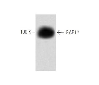
Date published: 2026-1-8

1-800-457-3801

SCBT Portrait Logo
Seach Input

GAP1m Antibody (15): sc-135916

4.0(2)
Write a reviewAsk a question

Datasheets
  • GAP1m Antibody (15) is a mouse monoclonal IgG1 κ provided at 200 µg/ml
  • raised against amino acids 20-200 of GAP1m of rat origin
  • recommended for detection of GAP1m of mouse, rat and human origin by WB, IP and IF; not recommended for IP
  • m-IgG Fc BP-HRP is the preferred secondary detection reagent for GAP1m Antibody (15) for WB applications. This reagent is now offered in a bundle with GAP1m Antibody (15) (see ordering information below).

    QUICK LINKS

    SEE ALSO...

    GAP1m Antibody (15) is a mouse monoclonal IgG1 kappa light chain antibody that detects GAP1m protein of mouse, rat, and human origin by western blotting (WB), immunoprecipitation (IP), and immunofluorescence (IF). Anti-GAP1m antibody (15) is available as the non-conjugated format. GAP1m protein plays a crucial role in regulating the activity of Ras p21, a key player in cell signaling pathways that control cell growth and differentiation. By facilitating the hydrolysis of GTP bound to Ras p21, GAP1m enhances the conversion of Ras to its inactive GDP-bound form, thereby acting as a critical regulator of cellular signaling. This function is particularly important in preventing uncontrolled cell proliferation, which can lead to cancer. GAP1m is predominantly expressed in the brain, placenta, and kidney, highlighting its significance in various physiological processes. GAP1m contains a GAP catalytic domain, a phospholipid-binding region, and a domain homologous to a unique region of Bruton′s tyrosine kinase (Btk), which underscores its multifaceted role in cellular signaling and interaction with other proteins.

    For Research Use Only. Not Intended for Diagnostic or Therapeutic Use.

    Alexa Fluor® is a trademark of Molecular Probes Inc., OR., USA

    LI-COR® and Odyssey® are registered trademarks of LI-COR Biosciences

    GAP1m Antibody (15) References:

    1. Molecular cloning of cDNAs encoding the GAP-associated protein p190: implications for a signaling pathway from ras to the nucleus.  |  Settleman, J., et al. 1992. Cell. 69: 539-49. PMID: 1581965
    2. The GTPase superfamily: a conserved switch for diverse cell functions.  |  Bourne, HR., et al. 1990. Nature. 348: 125-32. PMID: 2122258
    3. A guide to low molecular weight GTPases.  |  Sanders, DA. 1990. Cell Growth Differ. 1: 251-8. PMID: 2150754
    4. A cytoplasmic protein stimulates normal N-ras p21 GTPase, but does not affect oncogenic mutants.  |  Trahey, M. and McCormick, F. 1987. Science. 238: 542-5. PMID: 2821624
    5. ras genes.  |  Barbacid, M. 1987. Annu Rev Biochem. 56: 779-827. PMID: 3304147
    6. A novel mammalian Ras GTPase-activating protein which has phospholipid-binding and Btk homology regions.  |  Maekawa, M., et al. 1994. Mol Cell Biol. 14: 6879-85. PMID: 7935405

    Ordering Information

    Product NameCatalog #UNITPriceQtyFAVORITES

    GAP1m Antibody (15)

    sc-135916
    200 µg/ml
    $322.00

    GAP1m Antibody (15): m-IgG Fc BP-HRP Bundle

    sc-551135
    200 µg Ab; 10 µg BP
    $361.00

    What is the recommended fixation method for immunofluorescence staining with GAP1m (15): sc-135916 monoclonal antibody?

    Asked by: jenniferc
    Thank you for your inquiry. We recommend fixing cells for 5 minutes in -10ºC methanol and allowing to air dry. The full protocol can be viewed here: https://www.scbt.com/scbt/resources/protocols/immunofluorescence-cell-staining
    Answered by: Technical Support
    Date published: 2017-03-24
    • y_2025, m_12, d_30, h_8CST
    • bvseo_bulk, prod_bvqa, vn_bulk_3.0.42
    • cp_1, bvpage1
    • co_hasquestionsanswers, tq_1
    • loc_en_US, sid_135916, prod, sort_[SortEntry(order=LAST_APPROVED_ANSWER_SUBMISSION_TIME, direction=DESCENDING)]
    • clientName_scbt
    • bvseo_sdk, java_sdk, bvseo-4.0.0
    • CLOUD, getContent, 118ms
    • QUESTIONS, PRODUCT
    Rated 4 out of 5 by from Wonderful immunofluorescence membrane stainingWonderful immunofluorescence membrane staining in FHS cells. -SCBT QC
    Date published: 2015-03-27
    • y_2025, m_12, d_30, h_8
    • bvseo_bulk, prod_bvrr, vn_bulk_3.0.42
    • cp_1, bvpage1
    • co_hasreviews, tv_1, tr_1
    • loc_en_US, sid_135916, prod, sort_[SortEntry(order=SUBMISSION_TIME, direction=DESCENDING)]
    • clientName_scbt
    • bvseo_sdk, java_sdk, bvseo-4.0.0
    • CLOUD, getReviews, 17ms
    • REVIEWS, PRODUCT
    GAP1 m Antibody (15) is rated 4.0 out of 5 by 2.
    • y_2025, m_12, d_30, h_8
    • bvseo_bulk, prod_bvrr, vn_bulk_3.0.42
    • cp_1, bvpage1
    • co_hasreviews, tv_1, tr_1
    • loc_en_US, sid_135916, prod, sort_[SortEntry(order=SUBMISSION_TIME, direction=DESCENDING)]
    • clientName_scbt
    • bvseo_sdk, java_sdk, bvseo-4.0.0
    • CLOUD, getAggregateRating, 109ms
    • REVIEWS, PRODUCT