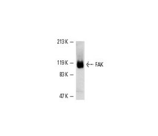
Date published: 2025-10-9

1-800-457-3801

SCBT Portrait Logo
Seach Input

FAK Antibody (H-1): sc-1688

4.5(12)
Write a reviewAsk a question

Datasheets
  • FAK Antibody (H-1) is a mouse monoclonal IgG1 κ, cited in 111 publications, provided at 200 µg/ml
  • raised against amino acids 903-1052 of FAK of mouse origin
  • Anti-FAK Antibody (H-1) is recommended for detection of FAK p125 of mouse, rat and human origin by WB, IP, IF, IHC(P) and FCM
  • FAK Antibody (H-1) is available conjugated to phycoerythrin for IF, IHC(P) and FCM
  • also available conjugated to Alexa Fluor® 594 for IF, IHC(P) and FCM
  • also available conjugated to Alexa Fluor® 680 or Alexa Fluor® 790 for WB (NIR), IF and FCM; for use with Near-Infrared (NIR) detection systems, such as LI-COR®Odyssey®, iBright™ FL1000, FluorChem™, Typhoon, Azure and other comparable systems
  • m-IgG Fc BP-HRP and m-IgG1 BP-HRP are the preferred secondary detection reagents for FAK Antibody (H-1) for WB and IHC(P) applications. These reagents are now offered in bundles with FAK Antibody (H-1) (see ordering information below).

QUICK LINKS

SEE ALSO...

FAK Antibody (H-1) is a mouse monoclonal IgG1 kappa light chain antibody that detects FAK p125 in mouse, rat, and human samples through applications such as western blotting (WB), immunoprecipitation (IP), immunofluorescence (IF), immunohistochemistry, and flow cytometry (FCM). FAK (H-1) antibody is available in both conjugated and non-conjugated forms, providing versatility for various experimental needs. Focal adhesion kinase (FAK) plays a crucial role in cellular signaling and is primarily located at focal adhesions, which are specialized structures that mediate the attachment of cells to the extracellular matrix. This localization is vital as FAK integrates signals from the extracellular environment, influencing cell migration, proliferation, and survival. FAK is concentrated at the basal edge of migrating keratinocytes during wound healing, suggesting significant involvement in the reepithelialization process. FAK′s protein tyrosine kinase activity is upregulated in response to mitogenic stimuli, highlighting importance in cellular responses to growth factors and neurotransmitters. FAK p125′s unique structural organization, as a cytoplasmic protein tyrosine kinase, distinguishes FAK from other kinases, underscoring specialized functions in cellular dynamics and tissue repair.

For Research Use Only. Not Intended for Diagnostic or Therapeutic Use.

Alexa Fluor® is a trademark of Molecular Probes Inc., OR., USA

LI-COR® and Odyssey® are registered trademarks of LI-COR Biosciences

FAK Antibody (H-1) References:

  1. Activation of transforming G protein-coupled receptors induces rapid tyrosine phosphorylation of cellular proteins, including p125FAK and the p130 v-src substrate.  |  Gutkind, JS. and Robbins, KC. 1992. Biochem Biophys Res Commun. 188: 155-61. PMID: 1329743
  2. Regulation of focal adhesion-associated protein tyrosine kinase by both cellular adhesion and oncogenic transformation.  |  Guan, JL. and Shalloway, D. 1992. Nature. 358: 690-2. PMID: 1379699
  3. Bombesin, vasopressin, and endothelin stimulation of tyrosine phosphorylation in Swiss 3T3 cells. Identification of a novel tyrosine kinase as a major substrate.  |  Zachary, I., et al. 1992. J Biol Chem. 267: 19031-4. PMID: 1382065
  4. Integrin-dependent phosphorylation and activation of the protein tyrosine kinase pp125FAK in platelets.  |  Lipfert, L., et al. 1992. J Cell Biol. 119: 905-12. PMID: 1385445
  5. Focal adhesion protein-tyrosine kinase phosphorylated in response to cell attachment to fibronectin.  |  Hanks, SK., et al. 1992. Proc Natl Acad Sci U S A. 89: 8487-91. PMID: 1528852
  6. pp125FAK a structurally distinctive protein-tyrosine kinase associated with focal adhesions.  |  Schaller, MD., et al. 1992. Proc Natl Acad Sci U S A. 89: 5192-6. PMID: 1594631
  7. Autophosphorylation of the focal adhesion kinase, pp125FAK, directs SH2-dependent binding of pp60src.  |  Schaller, MD., et al. 1994. Mol Cell Biol. 14: 1680-8. PMID: 7509446
  8. Potential role for focal adhesion kinase in migrating and proliferating keratinocytes near epidermal wounds and in culture.  |  Gates, RE., et al. 1994. Cell Growth Differ. 5: 891-9. PMID: 7986754

Ordering Information

Product NameCatalog #UNITPriceQtyFAVORITES

FAK Antibody (H-1)

sc-1688
200 µg/ml
$316.00

FAK Antibody (H-1): m-IgG Fc BP-HRP Bundle

sc-526472
200 µg Ab; 10 µg BP
$354.00

FAK Antibody (H-1): m-IgG1 BP-HRP Bundle

sc-531845
200 µg Ab; 20 µg BP
$354.00

FAK Antibody (H-1) PE

sc-1688 PE
200 µg/ml
$343.00

FAK Antibody (H-1) Alexa Fluor® 546

sc-1688 AF546
200 µg/ml
$357.00

FAK Antibody (H-1) Alexa Fluor® 594

sc-1688 AF594
200 µg/ml
$357.00

FAK Antibody (H-1) Alexa Fluor® 680

sc-1688 AF680
200 µg/ml
$357.00

FAK Antibody (H-1) Alexa Fluor® 790

sc-1688 AF790
200 µg/ml
$357.00

Hello, I am using Anti Fak SC-1688 in WB, but I see another bands in 250, 75 and 50 kD. Why is it recognizing other proteins?

Asked by: marithearceo
Thank you for your question. Please contact our Technical Service Department for assistance in troubleshooting this antibody.
Answered by: Technical Support
Date published: 2019-01-29

Can I ask for a quote for the product----FAK Antibody (H-1): sc-1688 PE? Thanks.

Asked by: Jen Ming
Thank you for your question. Our pricing in Australian Dollars (AUD) for this PE conjugated antibody is available on our website. You can change the currency and language settings on our website by going to the top of the page and using the drop-down menus. Please contact our Customer Care Department to request a formal quote. Or you can contact one of our authorized distributors in Australia. Please use the link below to find all of the contact information to request a quote, or place an order. https://www.scbt.com/scbt/resources/distributors
Answered by: Technical Support
Date published: 2017-07-31

I am planning on performing some IHC. Do you have recommendations for doing this will only mouse monoclonal antibodies?

Asked by: Cweed
We suggest using directly conjugated primary antibodies. For immunostaining, consider using sc-271126 which is available conjugated to FITC, AF647, and AF488.
Answered by: Technical Support 3
Date published: 2017-01-11

I am studying bovine samples. Do you think sc-1688 will work for my experiment?

Asked by: Cweed
Please contact our Technical Service Department and we would be happy to perform sequence analysis and review our data to answer any questions you may have about additional species reactivity.
Answered by: Technical Support 3
Date published: 2017-01-11
  • y_2025, m_9, d_24, h_6CST
  • bvseo_bulk, prod_bvqa, vn_bulk_3.0.42
  • cp_1, bvpage1
  • co_hasquestionsanswers, tq_4
  • loc_en_US, sid_1688, prod, sort_[SortEntry(order=LAST_APPROVED_ANSWER_SUBMISSION_TIME, direction=DESCENDING)]
  • clientName_scbt
  • bvseo_sdk, java_sdk, bvseo-4.0.0
  • CLOUD, getContent, 87ms
  • QUESTIONS, PRODUCT
Rated 4 out of 5 by from Work for Western blotUsing hASMC extract, detect a band in 120 kD and a stronger band in 50 kD.
Date published: 2017-11-12
Rated 5 out of 5 by from Great monoclonalStrong western blot and IHC data; low background !!
Date published: 2017-01-13
Rated 5 out of 5 by from Good for multiple applicationsGood antibody for flow cytometry and immunohistochemistry. Clear bands in Western blots as well, and the antibody is well-cited.
Date published: 2016-12-20
Rated 3 out of 5 by from Very good specificity but very low sensitivityVery good specificity but very low sensitivity for WB with samples from a rat cell line.
Date published: 2016-06-28
Rated 5 out of 5 by from This has worked well in WBThis has worked well in WB.
Date published: 2015-10-22
Rated 5 out of 5 by from Publishable data in IHC(P) using breast tumorPublishable data in IHC(P) using breast tumor, cervical epithelium and cervical carcinoma. -SCBT Publication Review
Date published: 2015-05-19
Rated 4 out of 5 by from Worked with transfected lysate by WBWorked with transfected lysate by WB -SCBT QC
Date published: 2015-03-17
Rated 4 out of 5 by from Robust band detected in WB using HeLa wholeRobust band detected in WB using HeLa whole cell lysate. -SCBT QC
Date published: 2015-01-29
  • y_2025, m_9, d_24, h_6
  • bvseo_bulk, prod_bvrr, vn_bulk_3.0.42
  • cp_1, bvpage1
  • co_hasreviews, tv_0, tr_12
  • loc_en_US, sid_1688, prod, sort_[SortEntry(order=SUBMISSION_TIME, direction=DESCENDING)]
  • clientName_scbt
  • bvseo_sdk, java_sdk, bvseo-4.0.0
  • CLOUD, getReviews, 9ms
  • REVIEWS, PRODUCT
FAK Antibody (H-1) is rated 4.5 out of 5 by 12.
  • y_2025, m_9, d_24, h_6
  • bvseo_bulk, prod_bvrr, vn_bulk_3.0.42
  • cp_1, bvpage1
  • co_hasreviews, tv_0, tr_12
  • loc_en_US, sid_1688, prod, sort_[SortEntry(order=SUBMISSION_TIME, direction=DESCENDING)]
  • clientName_scbt
  • bvseo_sdk, java_sdk, bvseo-4.0.0
  • CLOUD, getAggregateRating, 125ms
  • REVIEWS, PRODUCT