")
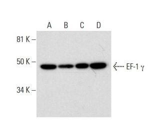
EF-1 γ Antibody (C-7): sc-393378
- EF-1 γ Antibody (C-7) is a mouse monoclonal IgG1 κ EF-1 γ antibody, cited in 2 publications, provided at 200 µg/ml
- raised against amino acids 204-384 mapping near the C-terminus of EF-1 γ of human origin
- EF-1 gamma Antibody (C-7) is recommended for detection of EF-1 γ of mouse, rat and human origin by WB, IP, IF, IHC(P) and ELISA
- Anti-EF-1 gamma Antibody (C-7) is available conjugated to agarose for IP; HRP for WB, IHC(P) and ELISA; and to either phycoerythrin or FITC for IF, IHC(P) and FCM
- also available conjugated to Alexa Fluor® 488, Alexa Fluor® 546, Alexa Fluor® 594 or Alexa Fluor® 647 for WB (RGB), IF, IHC(P) and FCM, and for use with RGB fluorescent imaging systems, such as iBright™ FL1000, FluorChem™, Typhoon, Azure and other comparable systems
- also available conjugated to Alexa Fluor® 680 or Alexa Fluor® 790 for WB (NIR), IF and FCM; for use with Near-Infrared (NIR) detection systems, such as LI-COR®Odyssey®, iBright™ FL1000, FluorChem™, Typhoon, Azure and other comparable systems
- m-IgG Fc BP-HRP is the preferred secondary detection reagent for EF-1 γ Antibody (C-7) for WB and IHC(P) applications. This reagent is now offered in a bundle with EF-1 γ Antibody (C-7) (see ordering information below).

EF-1 γ Antibody (C-7) is a mouse monoclonal IgG1 kappa light chain antibody that detects EF-1 gamma protein of mouse, rat, and human origin by western blotting (WB), immunoprecipitation (IP), immunofluorescence (IF), immunohistochemistry, and enzyme-linked immunosorbent assay (ELISA). EF-1 γ (C-7) antibody is available in both non-conjugated and various conjugated forms, including agarose, horseradish peroxidase (HRP), phycoerythrin (PE), fluorescein isothiocyanate (FITC), and multiple Alexa Fluor® conjugates. EF-1 gamma protein, also known as EEF1G or GIG35, is a crucial 437 amino acid subunit of the elongation factor-1 complex, which is essential for delivering aminoacyl-tRNAs to the ribosome during protein synthesis. EF-1 gamma protein is predominantly expressed in various tissues, including stomach, pancreas, brain, lung, kidney, intestine, liver, and spleen, and features an N-terminal glutathione transferase domain that anchors the complex to different cellular components. EF-1 gamma protein is implicated in assembling multiprotein complexes that include aminoacyl-tRNA synthetases, which are vital for accurate protein translation. Increased expression levels of EF-1 gamma protein have been linked to pancreatic cancer, indicating potential involvement in oncogenic processes and making EF-1 gamma protein a significant target for cancer research.
Alexa Fluor® is a trademark of Molecular Probes Inc., OR., USA
LI-COR® and Odyssey® are registered trademarks of LI-COR Biosciences
EF-1 γ Antibody (C-7) References:
- Expression of elongation factor-1 gamma-related sequence in human pancreatic cancer. | Lew, Y., et al. 1992. Pancreas. 7: 144-52. PMID: 1372736
- Elongation factor-1 messenger-RNA levels in cultured cells are high compared to tissue and are not drastically affected further by oncogenic transformation. | Sanders, J., et al. 1992. Nucleic Acids Res. 20: 5907-10. PMID: 1461723
- Molecular hierarchy in neurons differentiated from mouse ES cells containing a single human chromosome 21. | Wang, CC., et al. 2004. Biochem Biophys Res Commun. 314: 335-50. PMID: 14733910
- Gene expression profiling of human HBV- and/or HCV-associated hepatocellular carcinoma cells using expressed sequence tags. | Yoon, SY., et al. 2006. Int J Oncol. 29: 315-27. PMID: 16820872
- Temporal expression of transcripts related to embryo quality in bovine embryos cultured from the two-cell to blastocyst stage in vitro or in vivo. | Corcoran, D., et al. 2007. Mol Reprod Dev. 74: 972-7. PMID: 17219429
- Eukaryotic translation elongation factor 1 gamma contains a glutathione transferase domain--study of a diverse, ancient protein superfamily using motif search and structural modeling. | Koonin, EV., et al. 1994. Protein Sci. 3: 2045-54. PMID: 7703850
Ordering Information
| Product Name | Catalog # | UNIT | Price | Qty | FAVORITES | |
EF-1 γ Antibody (C-7) | sc-393378 | 200 µg/ml | $322.00 | |||
EF-1 γ Antibody (C-7): m-IgG Fc BP-HRP Bundle | sc-526244 | 200 µg Ab; 10 µg BP | $361.00 | |||
EF-1 γ Antibody (C-7) AC | sc-393378 AC | 500 µg/ml, 25% agarose | $424.00 | |||
EF-1 γ Antibody (C-7) HRP | sc-393378 HRP | 200 µg/ml | $322.00 | |||
EF-1 γ Antibody (C-7) FITC | sc-393378 FITC | 200 µg/ml | $336.00 | |||
EF-1 γ Antibody (C-7) PE | sc-393378 PE | 200 µg/ml | $349.00 | |||
EF-1 γ Antibody (C-7) Alexa Fluor® 488 | sc-393378 AF488 | 200 µg/ml | $364.00 | |||
EF-1 γ Antibody (C-7) Alexa Fluor® 546 | sc-393378 AF546 | 200 µg/ml | $364.00 | |||
EF-1 γ Antibody (C-7) Alexa Fluor® 594 | sc-393378 AF594 | 200 µg/ml | $364.00 | |||
EF-1 γ Antibody (C-7) Alexa Fluor® 647 | sc-393378 AF647 | 200 µg/ml | $364.00 | |||
EF-1 γ Antibody (C-7) Alexa Fluor® 680 | sc-393378 AF680 | 200 µg/ml | $364.00 | |||
EF-1 γ Antibody (C-7) Alexa Fluor® 790 | sc-393378 AF790 | 200 µg/ml | $364.00 |