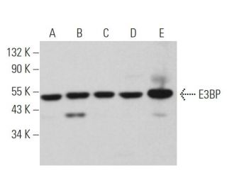
Date published: 2026-2-14

1-800-457-3801

SCBT Portrait Logo
Seach Input

E3BP Antibody (H-6): sc-377255

5.0(3)
Write a reviewAsk a question

Datasheets
  • E3BP Antibody (H-6) is a mouse monoclonal IgG2b κ E3BP antibody, cited in 3 publications, provided at 200 µg/ml
  • raised against amino acids 151-280 mapping within an internal region of E3BP of human origin
  • E3BP Antibody (H-6) is recommended for detection of E3BP of mouse, rat and human origin by WB, IP, IF, IHC(P) and ELISA
  • Anti-E3BP Antibody (H-6) is available conjugated to agarose for IP; HRP for WB, IHC(P) and ELISA; and to either phycoerythrin or FITC for IF, IHC(P) and FCM
  • also available conjugated to Alexa Fluor® 488, Alexa Fluor® 546, Alexa Fluor® 594 or Alexa Fluor® 647 for WB (RGB), IF, IHC(P) and FCM, and for use with RGB fluorescent imaging systems, such as iBright™ FL1000, FluorChem™, Typhoon, Azure and other comparable systems
  • also available conjugated to Alexa Fluor® 680 or Alexa Fluor® 790 for WB (NIR), IF and FCM; for use with Near-Infrared (NIR) detection systems, such as LI-COR®Odyssey®, iBright™ FL1000, FluorChem™, Typhoon, Azure and other comparable systems
  • m-IgGκ BP-HRP is the preferred secondary detection reagent for E3BP Antibody (H-6) for WB and IHC(P) applications. This reagent is now offered in a bundle with E3BP Antibody (H-6) (see ordering information below). For additional m-IgGκ BP conjugates see our complete list of Mouse IgG Binding Proteins.
Crispr Promo Banner

QUICK LINKS

SEE ALSO...

E3BP Antibody (H-6) is a mouse monoclonal IgG2b kappa light chain antibody that detects E3BP protein of mouse, rat, and human origin by western blotting (WB), immunoprecipitation (IP), immunofluorescence (IF), immunohistochemistry, and enzyme-linked immunosorbent assay (ELISA). Anti-E3BP antibody (H-6) is available in both non-conjugated and various conjugated forms, including agarose, horseradish peroxidase (HRP), phycoerythrin (PE), fluorescein isothiocyanate (FITC), and multiple Alexa Fluor® conjugates. E3BP protein, also known as PDHX (Pyruvate dehydrogenase protein X component), plays a crucial role in the pyruvate dehydrogenase (PDH) complex, which is essential for cellular metabolism as this complex serves as the primary link between glycolysis and the tricarboxylic acid (TCA) cycle by catalyzing the conversion of pyruvate into acetyl-CoA. E3BP is a 501 amino acid mitochondrial protein that is vital for anchoring the E3 component to the E2 core of the PDH complex, a process necessary for the complex′s functionality. Deficiencies in E3BP can lead to severe metabolic disorders, including lactic acidosis and developmental delays, highlighting the importance of E3BP in maintaining metabolic homeostasis. Clinical implications of E3BP deficiency can manifest as a range of neurological symptoms, including seizures and cerebellar ataxia, underscoring E3BP′s critical role in both energy metabolism and neurological health.

For Research Use Only. Not Intended for Diagnostic or Therapeutic Use.

Alexa Fluor® is a trademark of Molecular Probes Inc., OR., USA

LI-COR® and Odyssey® are registered trademarks of LI-COR Biosciences

E3BP Antibody (H-6) References:

  1. Mitochondrial dysfunction in a patient with Joubert syndrome.  |  Morava, E., et al. 2005. Neuropediatrics. 36: 214-7. PMID: 15944909
  2. Leigh's disease due to a new mutation in the PDHX gene.  |  Schiff, M., et al. 2006. Ann Neurol. 59: 709-14. PMID: 16566017
  3. PDC-E3BP is not a dominant T-cell autoantigen in primary biliary cirrhosis.  |  McHugh, A., et al. 2006. Liver Int. 26: 406-13. PMID: 16629643
  4. A new level of architectural complexity in the human pyruvate dehydrogenase complex.  |  Smolle, M., et al. 2006. J Biol Chem. 281: 19772-80. PMID: 16679318
  5. A novel gross deletion caused by non-homologous recombination of the PDHX gene in a patient with pyruvate dehydrogenase deficiency.  |  Miné, M., et al. 2006. Mol Genet Metab. 89: 106-10. PMID: 16843025
  6. Pyruvate dehydrogenase E3 binding protein (protein X) deficiency.  |  Brown, RM., et al. 2006. Dev Med Child Neurol. 48: 756-60. PMID: 16904023
  7. Subunit and catalytic component stoichiometries of an in vitro reconstituted human pyruvate dehydrogenase complex.  |  Brautigam, CA., et al. 2009. J Biol Chem. 284: 13086-98. PMID: 19240034
  8. Defects in the E2 lipoyl transacetylase and the X-lipoyl containing component of the pyruvate dehydrogenase complex in patients with lactic acidemia.  |  Robinson, BH., et al. 1990. J Clin Invest. 85: 1821-4. PMID: 2112155

Ordering Information

Product NameCatalog #UNITPriceQtyFAVORITES

E3BP Antibody (H-6)

sc-377255
200 µg/ml
$322.00

E3BP Antibody (H-6): m-IgGκ BP-HRP Bundle

sc-523250
200 µg Ab, 40 µg BP
$361.00

E3BP Antibody (H-6) AC

sc-377255 AC
500 µg/ml, 25% agarose
$424.00

E3BP Antibody (H-6) HRP

sc-377255 HRP
200 µg/ml
$322.00

E3BP Antibody (H-6) FITC

sc-377255 FITC
200 µg/ml
$336.00

E3BP Antibody (H-6) PE

sc-377255 PE
200 µg/ml
$349.00

E3BP Antibody (H-6) Alexa Fluor® 488

sc-377255 AF488
200 µg/ml
$364.00

E3BP Antibody (H-6) Alexa Fluor® 546

sc-377255 AF546
200 µg/ml
$364.00

E3BP Antibody (H-6) Alexa Fluor® 594

sc-377255 AF594
200 µg/ml
$364.00

E3BP Antibody (H-6) Alexa Fluor® 647

sc-377255 AF647
200 µg/ml
$364.00

E3BP Antibody (H-6) Alexa Fluor® 680

sc-377255 AF680
200 µg/ml
$364.00

E3BP Antibody (H-6) Alexa Fluor® 790

sc-377255 AF790
200 µg/ml
$364.00

Can E3BP (H-6): sc-377255 monoclonal antibody be used to stain formalin-fixed, paraffin-embedded (FFPE) tissue sections?

Asked by: Randy McDonald
Thank you for your question. Yes, E3BP (H-6): sc-377255 has been validated for use in IHC with paraffin-embedded sections. We recommend performing antigen retrieval with sodium citrate buffer (pH 6) and heat. The full protocol can be found here: https://www.scbt.com/scbt/resources/protocols/immunoperoxidase-staining
Answered by: Technical Support
Date published: 2017-02-25
  • y_2026, m_2, d_12, h_7CST
  • bvseo_bulk, prod_bvqa, vn_bulk_3.0.42
  • cp_1, bvpage1
  • co_hasquestionsanswers, tq_1
  • loc_en_US, sid_377255, prod, sort_[SortEntry(order=LAST_APPROVED_ANSWER_SUBMISSION_TIME, direction=DESCENDING)]
  • clientName_scbt
  • bvseo_sdk, java_sdk, bvseo-4.0.0
  • CLOUD, getContent, 90ms
  • QUESTIONS, PRODUCT
Rated 5 out of 5 by from E3BP (H-6)E3BP (H-6): sc. Near-infrared western blot analysis of LDH-A expression in MEF whole cell lysates. Blocked with 5% NFDM for 1 hour. Detection reagent used: DAM CW800 (Licor).
Date published: 2018-06-28
Rated 5 out of 5 by from Produced beautiful immunofluorescence mitochondrialProduced beautiful immunofluorescence mitochondrial staining in methanol-fixed HeLa cells. -SCBT QC
Date published: 2015-02-07
  • y_2026, m_2, d_12, h_7
  • bvseo_bulk, prod_bvrr, vn_bulk_3.0.42
  • cp_1, bvpage1
  • co_hasreviews, tv_1, tr_2
  • loc_en_US, sid_377255, prod, sort_[SortEntry(order=SUBMISSION_TIME, direction=DESCENDING)]
  • clientName_scbt
  • bvseo_sdk, java_sdk, bvseo-4.0.0
  • CLOUD, getReviews, 15ms
  • REVIEWS, PRODUCT
E3BP Antibody (H-6) is rated 5.0 out of 5 by 3.
  • y_2026, m_2, d_12, h_7
  • bvseo_bulk, prod_bvrr, vn_bulk_3.0.42
  • cp_1, bvpage1
  • co_hasreviews, tv_1, tr_2
  • loc_en_US, sid_377255, prod, sort_[SortEntry(order=SUBMISSION_TIME, direction=DESCENDING)]
  • clientName_scbt
  • bvseo_sdk, java_sdk, bvseo-4.0.0
  • CLOUD, getAggregateRating, 138ms
  • REVIEWS, PRODUCT