")
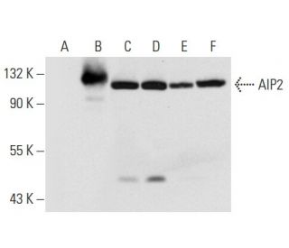
: sc-398090. Western blot analysis of AIP2 expression in non-transfected 293T: sc-117752 (A), human AIP2 transfected 293T: sc-116898 (B), K-562 (C), Jurkat (D), HeLa (E) and NIH/3T3 (F) whole cell lysates.")
: sc-398090. Western blot analysis of AIP2 expression in K-562 (A), A549 (B), MDA-MB-435S (C), EOC 20 (D), C6 (E) and L6 (F) whole cell lysates.")
AIP2 Antibody (A-3): sc-398090
- AIP2 Antibody (A-3) is a mouse monoclonal IgG2a κ AIP2 antibody, cited in 5 publications, provided at 200 µg/ml
- specific for an epitope mapping between amino acids 30-57 near the N-terminus of AIP2 of human origin
- AIP2 Antibody (A-3) is recommended for detection of AIP2 of mouse, rat and human origin by WB, IP, IF and ELISA
- Anti-AIP2 Antibody (A-3) is available conjugated to agarose for IP; HRP for WB, IHC(P) and ELISA; and to either phycoerythrin or FITC for IF, IHC(P) and FCM
- also available conjugated to Alexa Fluor® 488, Alexa Fluor® 546, Alexa Fluor® 594 or Alexa Fluor® 647 for WB (RGB), IF, IHC(P) and FCM, and for use with RGB fluorescent imaging systems, such as iBright™ FL1000, FluorChem™, Typhoon, Azure and other comparable systems
- also available conjugated to Alexa Fluor® 680 or Alexa Fluor® 790 for WB (NIR), IF and FCM; for use with Near-Infrared (NIR) detection systems, such as LI-COR®Odyssey®, iBright™ FL1000, FluorChem™, Typhoon, Azure and other comparable systems
- m-IgG Fc BP-HRP and m-IgG2a BP-HRP are the preferred secondary detection reagents for AIP2 Antibody (A-3) for WB applications. These reagents are now offered in bundles with AIP2 Antibody (A-3) (see ordering information below).

QUICK LINKS
AIP2 Antibody (A-3) is a mouse monoclonal IgG2a kappa light chain antibody that detects AIP2 protein of mouse, rat, and human origin by western blotting (WB), immunoprecipitation (IP), immunofluorescence (IF), and enzyme-linked immunosorbent assay (ELISA). AIP2 (A-3) antibody is available in both non-conjugated and various conjugated forms, including agarose, horseradish peroxidase (HRP), phycoerythrin (PE), fluorescein isothiocyanate (FITC), and multiple Alexa Fluor® conjugates. AIP2 plays a crucial role in cellular processes by interacting with atrophin-1, particularly in the context of polyglutamine diseases, which are linked to neurodegenerative disorders such as Huntington′s disease. AIP2 is characterized by four WW domains, which are essential for protein-protein interactions, allowing binding to other proteins involved in signaling pathways and cellular regulation. The presence of a HECT domain in AIP2 suggests involvement in ubiquitin ligase activity, which is vital for protein degradation and turnover, thereby maintaining cellular homeostasis. AIP2′s interaction with other atrophin interacting proteins, such as AIP1 and AIP3, further emphasizes importance in the modulation of cellular functions and potential implications in disease mechanisms.
Alexa Fluor® is a trademark of Molecular Probes Inc., OR., USA
LI-COR® and Odyssey® are registered trademarks of LI-COR Biosciences
AIP2 Antibody (A-3) References:
- Unique roles of microRNA140 and its host gene WWP2 in cartilage biology. | Nakamura, Y., et al. 2008. J Musculoskelet Neuronal Interact. 8: 321-2. PMID: 19147957
- Enzymatic analysis of WWP2 E3 ubiquitin ligase using protein microarrays identifies autophagy-related substrates. | Jiang, H., et al. 2022. J Biol Chem. 298: 101854. PMID: 35331737
- LAPTM5 mediates immature B cell apoptosis and B cell tolerance by regulating the WWP2-PTEN-AKT pathway. | Wang, Y., et al. 2022. Proc Natl Acad Sci U S A. 119: e2205629119. PMID: 36037365
- Activation of E3 ubiquitin ligase WWP2 by non-receptor tyrosine kinase ACK1. | Zhu, J., et al. 2023. IUBMB Life. 75: 595-608. PMID: 36773333
- The role of WWP1 and WWP2 in bone/cartilage development and diseases. | Wang, Y., et al. 2024. Mol Cell Biochem. 479: 2907-2919. PMID: 38252355
- WWP2 deletion aggravates acute kidney injury by targeting CDC20/autophagy axis. | You, R., et al. 2024. J Adv Res.. PMID: 38909885
- WWP, a new amino acid motif present in single or multiple copies in various proteins including dystrophin and the SH3-binding Yes-associated protein YAP65. | André, B. and Springael, JY. 1994. Biochem Biophys Res Commun. 205: 1201-5. PMID: 7802651
- The rsp5-domain is shared by proteins of diverse functions. | Hofmann, K. and Bucher, P. 1995. FEBS Lett. 358: 153-7. PMID: 7828727
- The WW domain: a signalling site in dystrophin? | Bork, P. and Sudol, M. 1994. Trends Biochem Sci. 19: 531-3. PMID: 7846762
- Identification of novel human WW domain-containing proteins by cloning of ligand targets. | Pirozzi, G., et al. 1997. J Biol Chem. 272: 14611-6. PMID: 9169421
- The itchy locus encodes a novel ubiquitin protein ligase that is disrupted in a18H mice. | Perry, WL., et al. 1998. Nat Genet. 18: 143-6. PMID: 9462742
- Atrophin-1, the DRPLA gene product, interacts with two families of WW domain-containing proteins. | Wood, JD., et al. 1998. Mol Cell Neurosci. 11: 149-60. PMID: 9647693
Ordering Information
| Product Name | Catalog # | UNIT | Price | Qty | FAVORITES | |
AIP2 Antibody (A-3) | sc-398090 | 200 µg/ml | $322.00 | |||
AIP2 Antibody (A-3): m-IgG Fc BP-HRP Bundle | sc-526285 | 200 µg Ab; 10 µg BP | $361.00 | |||
AIP2 Antibody (A-3): m-IgG2a BP-HRP Bundle | sc-546561 | 200 µg Ab; 10 µg BP | $361.00 | |||
AIP2 Antibody (A-3) AC | sc-398090 AC | 500 µg/ml, 25% agarose | $424.00 | |||
AIP2 Antibody (A-3) HRP | sc-398090 HRP | 200 µg/ml | $322.00 | |||
AIP2 Antibody (A-3) FITC | sc-398090 FITC | 200 µg/ml | $336.00 | |||
AIP2 Antibody (A-3) PE | sc-398090 PE | 200 µg/ml | $349.00 | |||
AIP2 Antibody (A-3) Alexa Fluor® 488 | sc-398090 AF488 | 200 µg/ml | $364.00 | |||
AIP2 Antibody (A-3) Alexa Fluor® 546 | sc-398090 AF546 | 200 µg/ml | $364.00 | |||
AIP2 Antibody (A-3) Alexa Fluor® 594 | sc-398090 AF594 | 200 µg/ml | $364.00 | |||
AIP2 Antibody (A-3) Alexa Fluor® 647 | sc-398090 AF647 | 200 µg/ml | $364.00 | |||
AIP2 Antibody (A-3) Alexa Fluor® 680 | sc-398090 AF680 | 200 µg/ml | $364.00 | |||
AIP2 Antibody (A-3) Alexa Fluor® 790 | sc-398090 AF790 | 200 µg/ml | $364.00 | |||
AIP2 (A-3) Neutralizing Peptide | sc-398090 P | 100 µg/0.5 ml | $69.00 |