")
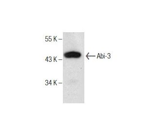
Abi-3 Anticorpo (C-7): sc-376982
- Abi-3 Anticorpo C-7é um anticorpo monoclonal produzido em camundongo IgG2a (kappa light chain) Abi-3 anticorpo fornecido em 200 µg/ml
- específico para um epítopo mapeado entre os aminoácidos 145-179 dentro de uma região interna de Abi-3 de human origem
- Abi-3 Anticorpo (C-7) é recomendado para a detecção de Abi-3 de mouse, rat e human origem em WB, IP, IF, IHC(P) e ELISA; reage também com outras espécies, incluindo bovine
- Anticorpo anti-Abi-3 O anticorpo anti-Abi-3 (C-7) está disponível conjugado com agarose para IP; HRP para WB, IHC(P) e ELISA; e com fioeritrina ou FITC para IF, IHC(P) e FCM
- também disponível conjugado com Alexa Fluor® 488, Alexa Fluor® 546, Alexa Fluor® 594 ou Alexa Fluor® 647 para WB (RGB), IF, IHC(P) e FCM, e para utilização com sistemas de imagiologia fluorescente RGB, tais como iBright™ FL1000, FluorChem™, Typhoon, Azure e outros sistemas comparáveis
- também disponível conjugado com Alexa Fluor® 680 ou Alexa Fluor® 790 para WB (NIR), IF e FCM; para utilização com sistemas de deteção de infravermelhos próximos (NIR), tais como LI-COR®Odyssey®, iBright™ FL1000, FluorChem™, Typhoon, Azure e outros sistemas comparáveis
- O m-IgGκ BP-HRP é o reagente de deteção secundário preferido para o anticorpo Abi-3 (C-7) para aplicações WB e IHC(P). Este reagente é agora oferecido num pacote com o anticorpo Abi-3 (C-7)(ver informações de encomenda abaixo). Para mais conjugados m-IgGκ BP, ver a nossa lista completa de Proteínas de Ligação IgG de Rato.

LINKS RÁPIDOS
O anticorpo Abi-3 (C-7) é um anticorpo monoclonal Abi-3 IgG2a κ de camundongo (também designado anticorpo Abi-3) que detecta a proteína Abi-3 de origem de camundongo, rato e humana por WB, IP, IF, IHC(P) e ELISA. O anticorpo Abi-3 (C-7) está disponível na forma de anticorpo anti-Abi-3 não conjugado, bem como em várias formas conjugadas de anticorpo anti-Abi-3, incluindo agarose, HRP, PE, FITC e vários conjugados Alexa Fluor®. O oncogene Abelson foi inicialmente identificado como o componente de transformação viral do vírus da leucemia murina de Abelson (A-MuLV). O gene Abelson (ABL1) codifica uma tirosina quinase com domínio SH2 que conduz a sinalização mitogénica de acordo com a ligação do recetor do fator de crescimento. As proteínas Abl interactor, Abi-1, Abi-2 e Abi-3, são proteínas com domínio SH3 que se ligam aos motivos ricos em prolina da Abl e activam a função cinase. Pensa-se que os membros da família Abi regulam negativamente o crescimento e a transformação celular, incluindo a transformação celular através de v-Abl, bem como medeiam a motilidade celular regulando a polimerização da actina nos lamelípodos e filópodos. A Abi-3, também designada por NESH, é uma proteína de 366 aminoácidos que interage com o TARSH, um gene relacionado com a senescência celular que actua como um fator de desencadeamento do desenvolvimento tumoral.
Alexa Fluor® é uma marca comercial da Molecular Probes Inc., OR., EUA
LI-COR® e Odyssey® são marcas registadas da LI-COR Biosciences
Referencias do Abi-3 Anticorpo (C-7):
- Isolamento e caraterização de um novo gene humano (NESH) que codifica uma molécula de sinalização putativa semelhante à proteína e3B1. | Miyazaki, K., et al. 2000. Biochim Biophys Acta. 1493: 237-41. PMID: 10978530
- A expressão forçada de NESH suprime a motilidade e a disseminação metastática de células malignas. | Ichigotani, Y., et al. 2002. Cancer Res. 62: 2215-9. PMID: 11956071
- Em busca de uma função para a família E3B1/Abi2/Argbp1/NESH (Revisão). | Ichigotani, Y., et al. 2002. Int J Mol Med. 9: 591-5. PMID: 12011975
- NESH (Abi-3) está presente no complexo Abi/WAVE mas não promove a fosforilação mediada por c-Abl. | Hirao, N., et al. 2006. FEBS Lett. 580: 6464-70. PMID: 17101133
- A proteína 53 do substrato do recetor de insulina (IRSp53) como parceiro de ligação da molécula antimetástase NESH, um membro da família de proteínas interactivas de Abelson. | Matsuda, S., et al. 2008. Ann Oncol. 19: 1356-1357. PMID: 18480067
- Implicação do gene relacionado com a senescência celular dependente de p53, TARSH, na supressão de tumores. | Wakoh, T., et al. 2009. Biochem Biophys Res Commun. 380: 807-12. PMID: 19338757
- A expressão da proteína NESH muda para o efeito adverso do mesilato de imatinib. | Matsuda, S., et al. 2008. Mol Oncol. 2: 16-9. PMID: 19383325
- Uma nova função de apoptose dependente de p53 de TARSH no desenvolvimento de tumores. | Wakoh, T., et al. 2009. Nagoya J Med Sci. 71: 109-14. PMID: 19994723
Informacoes sobre ordens
| Nome do Produto | Numero de Catalogo | UNID | Preco | Qde | FAVORITOS | |
Abi-3 Anticorpo (C-7) | sc-376982 | 200 µg/ml | $322.00 | |||
Pacote do Abi-3 (C-7): m-IgGκ BP-HRP | sc-552451 | 200 µg Ab; 40 µg BP | $361.00 | |||
Abi-3 Anticorpo (C-7) AC | sc-376982 AC | 500 µg/ml, 25% agarose | $424.00 | |||
Abi-3 Anticorpo (C-7) HRP | sc-376982 HRP | 200 µg/ml | $322.00 | |||
Abi-3 Anticorpo (C-7) FITC | sc-376982 FITC | 200 µg/ml | $336.00 | |||
Abi-3 Anticorpo (C-7) PE | sc-376982 PE | 200 µg/ml | $349.00 | |||
Abi-3 Anticorpo (C-7) Alexa Fluor® 488 | sc-376982 AF488 | 200 µg/ml | $364.00 | |||
Abi-3 Anticorpo (C-7) Alexa Fluor® 546 | sc-376982 AF546 | 200 µg/ml | $364.00 | |||
Abi-3 Anticorpo (C-7) Alexa Fluor® 594 | sc-376982 AF594 | 200 µg/ml | $364.00 | |||
Abi-3 Anticorpo (C-7) Alexa Fluor® 647 | sc-376982 AF647 | 200 µg/ml | $364.00 | |||
Abi-3 Anticorpo (C-7) Alexa Fluor® 680 | sc-376982 AF680 | 200 µg/ml | $364.00 | |||
Abi-3 Anticorpo (C-7) Alexa Fluor® 790 | sc-376982 AF790 | 200 µg/ml | $364.00 | |||
Abi-3 (C-7) peptídeo neutralizante | sc-376982 P | 100 µg/0.5 ml | $69.00 |