")
Anticorps VEGF-C (E-6): sc-374628
- L'anticorps VEGF-C E-6 est un monoclonal IgG2a κ L'Anticorps VEGF-C, cité dans 29 publications, fourni en 200 µg/ml
- spécifique d'un épitope situé entre les acides aminés 103-137 près de N-terminus de VEGF-C d'origine human
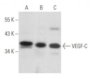
- L'Anticorps VEGF-C (E-6) est recommandé pour la détection de precursor and mature VEGF-C d'origine mouse, rat et human par WB, IP, IF, IHC(P) et ELISA
- Anti-L'Anticorps VEGF-C (E-6) est disponible conjugué à l'agarose pour IP; à l'HRP pour WB, IHC(P) et ELISA; et soit à la phycoerythrin ou FITC pour IF, IHC(P) et FCM
- aussi disponible conjugué à l'Alexa Fluor® 488, Alexa Fluor® 546, Alexa Fluor® 594 ou Alexa Fluor® 647 pour WB (RGB), IF, IHC(P) et FCM
- aussi disponible conjugué à l'Alexa Fluor® 680 ou Alexa Fluor® 790 pour WB (NIR), IF et FCM
- m-IgG Fc BP-HRP, 2a BP-HRP">m-IgG2a BP-HRP et m-IgGκ BP-HRP sont les réactifs de détection secondaire préférés pour l'anticorps VEGF-C (E-6) pour les applications WB et IHC(P). Ces réactifs sont désormais proposés en lots avec l'anticorps VEGF-C (E-6)(voir les informations de commande ci-dessous).

ACCÈS RAPIDE AUX LIENS
L'anticorps VEGF-C (E-6) est un anticorps monoclonal IgG2a κ de souris qui détecte la protéine VEGF-C de souris, de rat et d'homme par WB, IP, IF, IHC(P) et ELISA. L'anticorps VEGF-C (E-6) est disponible sous forme d'anticorps anti-VEGF-C non conjugué, ainsi que sous plusieurs formes conjuguées d'anticorps anti-VEGF-C, y compris agarose, HRP, PE, FITC et plusieurs conjugués Alexa Fluor®. L'angiogenèse est considérée comme un événement précoce dans la tumorigenèse et peut faciliter la progression de la tumeur et la formation de métastases. Plusieurs facteurs de croissance ayant une activité angiogénique ont été décrits. Il s'agit notamment du facteur de croissance des fibroblastes (FGF), du facteur de croissance dérivé des plaquettes (PDGF) et du facteur de croissance de l'endothélium vasculaire (VEGF). Le VEGF est une glycoprotéine dimérique dont la structure est homologue à celle du PDGF. Plusieurs variantes du VEGF ont été décrites et résultent d'un épissage alternatif de l'ARNm. On a supposé que le VEGF pouvait fonctionner comme un facteur d'angiogenèse tumorale in vivo. Deux autres protéines, appelées VEGF-B et VEGF-C, partagent un degré significatif d'homologie avec le VEGF. Le VEGF-B est abondamment exprimé dans le cœur et les muscles squelettiques et est fréquemment coexprimé avec le VEGF. Le VEGF-C se lie à Flt-4 et Flk-1 et les active spécifiquement. Les gènes qui codent pour le VEGF-B et le VEGF-C ont été localisés sur les chromosomes 11q13.1 et 4q34.3, respectivement.
Alexa Fluor® est une marque déposée de Molecular Probes Inc., OR., USA
LI-COR® et Odyssey® sont marques déposées de LI-COR Biosciences
Anticorps VEGF-C (E-6) Références:
- Le facteur de croissance de l'endothélium vasculaire est un facteur potentiel d'angiogenèse tumorale dans les gliomes humains in vivo. | Plate, KH., et al. 1992. Nature. 359: 845-8. PMID: 1279432
- Expression du facteur de croissance de l'endothélium vasculaire au cours de l'angiogenèse embryonnaire et de la différenciation des cellules endothéliales. | Breier, G., et al. 1992. Development. 114: 521-32. PMID: 1592003
- La famille des polypeptides du facteur de croissance de l'endothélium vasculaire. | Ferrara, N., et al. 1991. J Cell Biochem. 47: 211-8. PMID: 1791185
- Le VEGF-C régule de manière différentielle l'expression du VEGF-A dans les cellules oculaires et cancéreuses; il favorise l'angiogenèse par le biais de la voie RhoA. | Kumar, B., et al. 2011. Angiogenesis. 14: 371-80. PMID: 21698469
- Induction de l'angiogenèse pendant la transition de l'hyperplasie à la néoplasie. | Folkman, J., et al. 1989. Nature. 339: 58-61. PMID: 2469964
- Le VEGF-C est nécessaire à l'entretien des vaisseaux lymphatiques intestinaux et à l'absorption des lipides. | Nurmi, H., et al. 2015. EMBO Mol Med. 7: 1418-25. PMID: 26459520
- L'administration de VEGF-C à l'aide d'anticorps réduit efficacement l'inflammation chronique de la peau. | Schwager, S., et al. 2018. JCI Insight. 3: PMID: 30518687
- VEGF-C et mortalité chez les patients atteints d'une maladie coronarienne présumée ou avérée. | Wada, H., et al. 2018. J Am Heart Assoc. 7: e010355. PMID: 30554564
- Le VEGF-C associé aux vésicules extracellulaires favorise la lymphangiogenèse et l'infiltration des cellules immunitaires dans l'endométriose. | Li, WN., et al. 2020. Proc Natl Acad Sci U S A. 117: 25859-25868. PMID: 33004630
- L'administration artérielle de VEGF-C stabilise les lésions athérosclérotiques. | Silvestre-Roig, C., et al. 2021. Circ Res. 128: 284-286. PMID: 33210556
- Le VEGF-C contribue à la croissance tumorale et à la formation de métastases en favorisant la diaphonie entre l'EMT et les cellules épithéliales du cancer du sein. | Kong, D., et al. 2021. Oncogene. 40: 964-979. PMID: 33299122
- La simvastatine atténue les lésions du système lymphatique via la voie VEGF-C/VEGFR3/PI3K-Akt après une hémorragie intracérébrale expérimentale. | Liao, J., et al. 2024. Brain Res Bull. 216: 111045. PMID: 39097032
Informations pour la commande
| Nom du produit | Ref. Catalogue | COND. | Prix HT | QTÉ | Favoris | |
Anticorps VEGF-C (E-6) | sc-374628 | 200 µg/ml | $322.00 | |||
VEGF-C (E-6): m-IgG Fc BP-HRP Kit | sc-529785 | 200 µg Ab; 10 µg BP | $361.00 | |||
VEGF-C (E-6): m-IgGκ BP-HRP Kit | sc-522795 | 200 µg Ab, 40 µg BP | $361.00 | |||
VEGF-C (E-6): m-IgG2a BP-HRP Kit | sc-547378 | 200 µg Ab; 10 µg BP | $361.00 | |||
Anticorps VEGF-C (E-6) AC | sc-374628 AC | 500 µg/ml, 25% agarose | $424.00 | |||
Anticorps VEGF-C (E-6) HRP | sc-374628 HRP | 200 µg/ml | $322.00 | |||
Anticorps VEGF-C (E-6) FITC | sc-374628 FITC | 200 µg/ml | $336.00 | |||
Anticorps VEGF-C (E-6) PE | sc-374628 PE | 200 µg/ml | $349.00 | |||
Anticorps VEGF-C (E-6) Alexa Fluor® 488 | sc-374628 AF488 | 200 µg/ml | $364.00 | |||
Anticorps VEGF-C (E-6) Alexa Fluor® 546 | sc-374628 AF546 | 200 µg/ml | $364.00 | |||
Anticorps VEGF-C (E-6) Alexa Fluor® 594 | sc-374628 AF594 | 200 µg/ml | $364.00 | |||
Anticorps VEGF-C (E-6) Alexa Fluor® 647 | sc-374628 AF647 | 200 µg/ml | $364.00 | |||
Anticorps VEGF-C (E-6) Alexa Fluor® 680 | sc-374628 AF680 | 200 µg/ml | $364.00 | |||
Anticorps VEGF-C (E-6) Alexa Fluor® 790 | sc-374628 AF790 | 200 µg/ml | $364.00 | |||
VEGF-C (E-6) peptide neutralisant | sc-374628 P | 100 µg/0.5 ml | $69.00 |