")
pan-Cytokeratin Antikörper (AE1/AE3): sc-81714
- pan-Cytokeratin Antikörper (AE1/AE3) ist ein Maus monoklonales IgG1 κ, verwendet in 76 wissenschaftlichen Veröffentlichungen, in einer Menge von 200 µg/ml
- gegen epidermale Keratine human
- Empfohlen für die Detektion von most acidic (type I) and basic (type II) Cytokeratins aus der Spezies mouse, rat, human, bovine und rabbit per WB, IP, IF und IHC(P)
- erhältlich als Konjugat mit HRP für WB, IHC(P) und ELISA
- m-IgG Fc BP-HRP, 1 BP-HRP">m-IgG1 BP-HRP und m-IgGκ BP-HRP sind die bevorzugten sekundären Nachweisreagenzien für pan-Cytokeratin Antikörper (AE1/AE3) für WB- und IHC(P)-Anwendungen. Diese Reagenzien werden jetzt in Bündeln mit pan-Cytokeratin Antikörper (AE1/AE3) angeboten(siehe Bestellinformationen unten).

Direktverknüpfungen
Siehe auch...
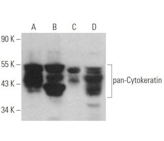
Der pan-Cytokeratin-Antikörper (AE1/AE3) ist ein monoklonaler IgG1-Kappa-Leichtketten-Antikörper der Maus, der pan-Cytokeratin in mehreren Spezies, darunter Maus, Ratte, Mensch, Rind und Kaninchen, durch Anwendungen wie Western Blot (WB), Immunop Immunopräzipitation (IP), Immunfluoreszenz (IF) und Immunhistochemie mit Paraffinschnitten (IHCP) nachgewiesen werden kann. Der anti-pan-Cytokeratin-Antikörper (AE1/AE3) ist sowohl in konjugierter als auch in nicht konjugierter Form erhältlich und bietet somit Flexibilität für verschiedene experimentelle Anforderungen. Cytokeratine, die vom monoklonalen pan-Cytokeratin-Antikörper (AE1/AE3) erkannt werden, sind eine wichtige Gruppe von Intermediärfilamentproteinen, die die strukturelle Integrität von Epithelzellen aufrechterhalten. Diese Proteine werden paarweise exprimiert und sind für die zelluläre Differenzierung und Gewebespezialisierung unerlässlich, was sie für die Krebsforschung besonders wertvoll macht. Bestimmte Zytokeratine dienen als zuverlässige Biomarker zur Unterscheidung zwischen verschiedenen Tumorarten; die Zytokeratine 10 und 13 sind häufig bei bestimmten Plattenepithelkarzinomen erhöht, während Zytokeratin 18 vorwiegend bei Adenokarzinomen und Basalzellkarzinomen exprimiert wird. Die Expressionsmuster von Zytokeratinen helfen bei der Charakterisierung bösartiger Tumore und geben Aufschluss über die Funktion und Pathologie von Epithelzellen.
Alexa Fluor® ist ein Markenzeichen von Molecular Probes Inc., OR., USA
LI-COR® und Odyssey® sind Markenzeichen von LI-COR Biosciences
Bestellinformation
| Produkt | Katalog # | EINHEIT | Preis | ANZAHL | Favoriten | |
pan-Cytokeratin Antikörper (AE1/AE3) | sc-81714 | 200 µg/ml | $322.00 | |||
pan-Cytokeratin (AE1/AE3): m-IgG Fc BP-HRP Bundle | sc-528712 | 200 µg Ab; 10 µg BP | $361.00 | |||
pan-Cytokeratin (AE1/AE3): m-IgGκ BP-HRP Bundle | sc-521181 | 200 µg Ab, 40 µg BP | $361.00 | |||
pan-Cytokeratin (AE1/AE3): m-IgG1 BP-HRP Bundle | sc-543082 | 200 µg Ab; 20 µg BP | $361.00 | |||
pan-Cytokeratin Antikörper (AE1/AE3) HRP | sc-81714 HRP | 200 µg/ml | $322.00 |