Date published: 2025-9-8

001 800-1338-3838

SCBT Portrait Logo
Seach Input

Occludin 항체 (E-5): sc-133256

4.2(37)
리뷰 쓰기질문하기

데이터 시트
  • Occludin 항체 E-5 는 마우스 monoclonal IgG2b κ Occludin 항체, 161간행물에 인용, 이며 200 µg/ml으로 제공합니다.
  • 아미노산 132-411에 대한 항체 human의 PI 3-키나아제의 Occludin의 SH2 도메인 내의 매핑
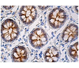
  • Occludin 항체 (E-5)는 WB, IP, IF, IHC(P) and ELISA으로 mouse, rat and human유래의 Occludin 를 감지하는 데에 추천한다.
  • 항-Occludin 항체(E-5)는 IP용 agarose와 결합되어 이용 가능하며, WB, IHC(P), ELISA용 HRP와 결합되어 이용 가능하며, IF, IHC(P), FCM용 phycoerythrin 또는 FITC와 결합되어 이용 가능합니다.
  • WB (RGB), IF, IHC(P) 와FCM, RGB fluorescent imaging systems, such as iBright™ FL1000, FluorChem™, Typhoon, Azure and other comparable systems에 사용가능한 Alexa Fluor® 488, Alexa Fluor® 546, Alexa Fluor® 594 or Alexa Fluor® 647결합제품도 있습니다.
  • WB (NIR), IF와FCM,Near-Infrared (NIR) detection systems, such as LI-COR®Odyssey®, iBright™ FL1000, FluorChem™, Typhoon, Azure and other comparable systems에 사용가능한 Alexa Fluor® 680 or Alexa Fluor® 790 결합제품도 있습니다.
  • Occludin (E-5): sc-133256무료10 µg 샘플을 신청하려면 기술지원팀 (또는 로컬 디스트리뷰터)에 연락주세요.
  • m-IgG2b BP-HRPm-IgGκ BP-HRP 는 Occludin 항체 (E-5) 가 WB and IHC(P) 에 응용될 때 우선으로 사용하는 이차 항체 검사 시약입니다.이 시약은Occludin 항체(E-5) 와 세트로 제공드립니다. (아래의 주문 정보를 참고해 주시길 바랍니다).

빠른 링크

더보기

Occludin 항체(E-5)는 마우스, 쥐 및 인간 유래의 Occludin 단백질을 WB, IP, IF, IHC(P) 및 ELISA로 검출하는 IgG2b κ 마우스 단일 클론 Occludin 항체(OCLN 항체로도 지정됨)입니다. 옥클루딘 항체(E-5)는 비접합 항-옥클루딘 항체 형태와 아가로스, HRP, PE, FITC 및 다중 Alexa Fluor® 접합체를 포함한 여러 접합 형태의 항-옥클루딘 항체 형태로 제공됩니다. 옥클루딘은 상피세포와 내피세포의 긴밀한 접합부와 밀접하게 관련된 필수 막 단백질입니다. Occludin은 상피와 내피의 타이트 접합(TJ) 구조에 있는 4면체 일체형 막 단백질로 두 개의 세포 외 고리를 포함할 수 있습니다. 이 단백질은 다양한 인산화 형태로 존재합니다. 인산화는 오클루딘의 국소화와 기능을 모두 조절하는 데 관여합니다. 오클루딘의 발현은 고도 불포화 지방산에 의해 상향 조절되어 내피 세포 저항성을 증가시키고 큰 분자에 대한 세포 투과성을 감소시킵니다. 오클루딘의 수준은 조직에 따라 크게 달라지며, 뇌 조직에서는 세포와 세포의 접촉 부위에서 오클루딘이 높게 발현됩니다. 비신경 조직에서는 발현이 낮고 불연속적인 분포를 보입니다. 상피 오클루딘의 상향 조절은 세포막 투과성을 향상시키는 역할을 할 수 있으며, 타이트 접합부의 손상과 관련이 있을 수 있습니다.

연구용으로만 사용가능합니다. 진단이나 치료용으로 사용불가합니다.

Alexa Fluor®는 미국 오리건주 Molecular Probes Inc.의 상표입니다.

LI-COR®와 Odyssey®는 LI-COR Biosciences의 등록 상표입니다.

Occludin 항체 (E-5) 참고문헌:

  1. 오클루딘의 분해는 뇌 미세혈관 내피 세포를 시험관 내 재관류 손상에 더 취약하게 만듭니다.  |  Zhang, Y., et al. 2021. J Neurochem. 156: 352-366. PMID: 32531803
  2. Occludin은 HIF-1α 신호 전달 경로를 통해 백반증에서 CD8+ T 세포와 멜라닌세포의 접착을 촉진합니다.  |  Zou, P., et al. 2022. Oxid Med Cell Longev. 2022: 6732972. PMID: 35222802
  3. 성공적인 재관류 후 혈청 오클루딘 수치가 높을수록 조기 신경학적 악화와 관련이 있다.  |  Li, W., et al. 2022. CNS Neurosci Ther. 28: 999-1007. PMID: 35338575
  4. 담배 연기에 의해 유발된 카텝신 S에 의한 오클루딘의 절단은 폐 상피 세포의 투과성을 증가시킨다.  |  Bigot, P., et al. 2022. Antioxidants (Basel). 12: PMID: 36670867
  5. 밀접 접합 단백질인 오클루딘은 쥐의 허혈성 뇌졸중 후 혈액-뇌 장벽의 완전성과 신경학적 기능을 조절합니다.  |  Sugiyama, S., et al. 2023. Sci Rep. 13: 2892. PMID: 36806348
  6. C형 간염 바이러스의 다단계 진입에 있어서 편광 간종양 구형체에 대한 표피 성장 인자 수용체, 클라우딘-1, 오클루딘의 역할.  |  So, CW., et al. 2023. PLoS Pathog. 19: e1011887. PMID: 38157366
  7. 오클루딘: 단단한 접합부에 국한되는 새로운 일체형 막 단백질.  |  Furuse, M., et al. 1993. J Cell Biol. 123: 1777-88. PMID: 8276896
  8. 타이트한 접합부의 분자 해부.  |  Tsukita, S., et al. 1996. Cell Struct Funct. 21: 381-5. PMID: 9118244
  9. 오클루딘의 인산화가 타이트 접합 형성에 관여할 가능성.  |  Sakakibara, A., et al. 1997. J Cell Biol. 137: 1393-401. PMID: 9182670
  10. 내피 세포에서 타이트 접합 투과성의 가능한 결정 인자로서의 오클루딘.  |  Hirase, T., et al. 1997. J Cell Sci. 110 (Pt 14): 1603-13. PMID: 9247194
  11. 오클루딘의 인산화는 오클루딘 국소화 및 타이트 접합부에서의 기능과 관련이 있습니다.  |  Wong, V. 1997. Am J Physiol. 273: C1859-67. PMID: 9435490
  12. 고도 불포화 지방산에 의한 타이트 접합 투과성 및 오클루딘 발현의 조절.  |  Jiang, WG., et al. 1998. Biochem Biophys Res Commun. 244: 414-20. PMID: 9514943

주문정보

제품명카탈로그 번호 단위가격수량관심품목

Occludin 항체 (E-5)

sc-133256
200 µg/ml
$316.00

Occludin (E-5): m-IgGκ BP-HRP 번들

sc-521292
200 µg Ab, 40 µg BP
$354.00

Occludin (E-5): m-IgG2b BP-HRP 번들

sc-548883
200 µg Ab; 10 µg BP
$354.00

Occludin 항체 (E-5) AC

sc-133256 AC
500 µg/ml, 25% agarose
$416.00

Occludin 항체 (E-5) HRP

sc-133256 HRP
200 µg/ml
$316.00

Occludin 항체 (E-5) FITC

sc-133256 FITC
200 µg/ml
$330.00

Occludin 항체 (E-5) PE

sc-133256 PE
200 µg/ml
$343.00

Occludin 항체 (E-5) Alexa Fluor® 488

sc-133256 AF488
200 µg/ml
$357.00

Occludin 항체 (E-5) Alexa Fluor® 546

sc-133256 AF546
200 µg/ml
$357.00

Occludin 항체 (E-5) Alexa Fluor® 594

sc-133256 AF594
200 µg/ml
$357.00

Occludin 항체 (E-5) Alexa Fluor® 647

sc-133256 AF647
200 µg/ml
$357.00

Occludin 항체 (E-5) Alexa Fluor® 680

sc-133256 AF680
200 µg/ml
$357.00

Occludin 항체 (E-5) Alexa Fluor® 790

sc-133256 AF790
200 µg/ml
$357.00

Que tipo de anticuerpo secundario es la alternativa para el uso de este anticuerpo primario. En el caso de IHC(P)

Asked by: Revrse
Que tipo de anticuerpo secundario es la alternativa para el uso de este anticuerpo primario. En el caso de IHC(P). https://www.scbt.com/p/m-igg2a-binding-protein-biotin https://www.scbt.com/p/m-igg2a-binding-protein-hrp
Answered by: BlakeJ
Date published: 2025-02-06

I used this antibody for IF,but it have some background,and I could not find the structure of occludin,could you give me some advice .

Asked by: Ning
Thank you for your question. Please contact our Technical Service Department, we are available at asia@scbio.cn, and happy to help you trouble-shooting.
Answered by: Technical Service SH
Date published: 2021-12-21

Am I amble to sample the antibody before I order it?

Asked by: jrauchut
Thank you for your question. Yes, for a limited time we are offering free 10 µg samples of this mouse monoclonal, Occludin Antibody (E-5): sc-133256. Please contact our Technical Service Department to request a free sample. Please call us toll-free at (800) 457-3801 option 2, e-mail <scbt@scbt.com> or use the live chat on our website.
Answered by: Technical Support
Date published: 2017-05-04

Can Occludin (E-5): sc-133256 monoclonal antibody be used to stain formalin-fixed, paraffin-embedded (FFPE) tissue sections?

Asked by: jenni
Thank you for your question. Yes, Occludin (E-5): sc-133256 monoclonal antibody has been validated for use in IHC with paraffin-embedded sections. We recommend performing antigen retrieval with sodium citrate buffer (pH 6) and heat. The full protocol can be found here: https://www.scbt.com/scbt/resources/protocols/immunoperoxidase-staining
Answered by: Technical Support 10
Date published: 2017-01-12

I am using the LiCor/Odyssey system. Which secondary antibody do you recommend using with Occludin (E-5): sc-133256?

Asked by: jenni
We recommend using one of our our exclusive Mouse IgG Binding Proteins (m-IgGκ) conjugated to CFL 680 or CFL 790. They are suitable for use with Near-Infrared (NIR) detection systems, such as LI-COR/Odyssey and other comparable systems. A complete list of available binding proteins is available on our website here: https://www.scbt.com/scbt/browse/support-products-mouse-igg-binding-proteins/_/N-ecrety
Answered by: Technical Support 10
Date published: 2017-01-12
  • y_2025, m_8, d_27, h_4CST
  • bvseo_bulk, prod_bvqa, vn_bulk_3.0.42
  • cp_1, bvpage1
  • co_hasquestionsanswers, tq_5
  • loc_ko_KR, sid_133256, prod, sort_[SortEntry(order=LAST_APPROVED_ANSWER_SUBMISSION_TIME, direction=DESCENDING)]
  • clientName_scbt
  • bvseo_sdk, java_sdk, bvseo-4.0.0
  • CLOUD, getContent, 114ms
  • QUESTIONS, PRODUCT
Rated 5 out of 5 by from Excellent antibodyI used anti-occludin (1:200) and worked perfectly in my breast cancer cell lysates (WB)
Date published: 2023-11-21
Rated 1 out of 5 by from Negative result in pig gut sampleAt necropsy, intestinal tract of pig were dissected. Sample was washed in NaCl 0.9% and post-fixed by immersion for 7 h in 10% buffered formalin (pH 7.4), dehydrated through a graded series of ethanol and finally embedded in Paraplast X-TRA. Four-µm thick sections were cut in the transversal plane and collected on silane-coated slides. After deparaffinization and rehydratation, the Avidin/Biotin blocking was performed and slides were then incubated overnight at room temperature in a dark moist chamber with anti-Occludin (E5) (sc-133256) diluted 1:100 in phosphate buffer saline (PBS). After washing in PBS, sections were incubated for 1.5 hour with Goat Anti-Mouse IgG Antibody (H+L), Biotinylated (6 μg/ml, Vector Laboratories Inc., Burlingame, CA, USA) in a dark moist chamber. The sections were washed twice in PBS and treated with Fluorescein–Avidin D (Vector Laboratories Inc., Burlingame, CA, USA). Finally, the slide with tissue sections was embedded in Vectashield Mounting Medium with DAPI (H-1200, Vector Laboratories Inc., Burlingame, CA, USA) and observed using a Confocal Laser Scanning Microscope (FluoView FV300, Olympus, Tokyo, Japan). Our results did not demonstrate any Occludine-immunopositivity in the epithelial cells of the pig gut mucosa, as expected.
Date published: 2022-12-15
Rated 1 out of 5 by from Negative result in pig gut sampleAt necropsy, intestinal tract of pig were dissected. Sample was washed in NaCl 0.9% and post-fixed by immersion for 7 h in 10% buffered formalin (pH 7.4), dehydrated through a graded series of ethanol and finally embedded in Paraplast X-TRA. Four-µm thick sections were cut in the transversal plane and collected on silane-coated slides. After deparaffinization and rehydratation, the Avidin/Biotin blocking was performed and slides were then incubated overnight at room temperature in a dark moist chamber with anti-Occludin (E5) (sc-133256) diluted 1:100 in phosphate buffer saline (PBS). After washing in PBS, sections were incubated for 1.5 hour with Goat Anti-Mouse IgG Antibody (H+L), Biotinylated (6 μg/ml, Vector Laboratories Inc., Burlingame, CA, USA) in a dark moist chamber. The sections were washed twice in PBS and treated with Fluorescein–Avidin D (Vector Laboratories Inc., Burlingame, CA, USA). Finally, the slide with tissue sections was embedded in Vectashield Mounting Medium with DAPI (H-1200, Vector Laboratories Inc., Burlingame, CA, USA) and observed using a Confocal Laser Scanning Microscope (FluoView FV300, Olympus, Tokyo, Japan). Our results did not demonstrate any Occludine-immunopositivity in the epithelial cells of the pig gut mucosa, as expected.
Date published: 2022-12-14
Rated 1 out of 5 by from Negative result in chickens intestinal sampleI tested this anti-occludin antibody on chicken ileum sample by immunohistochemistry. Antigen retrieval was performed using EDTA buffer (pH 9). Antibody was diluted 1:125 in 1,5% horse serum PBS and incubated 2h at RT. Antibody signal was detected using the avidin-biotin-proxidase method. However, it didn't work and no signal was shown.
Date published: 2022-03-17
Rated 4 out of 5 by from Good antibodyThe antibody works well for IHC staining performed on murine paraffin-embedded tissues. Specific and clear signal, no background. Highly recommended.
Date published: 2021-07-14
Rated 5 out of 5 by from Excelente resultadoCompre este producto hace días y es increíble el resultado
Date published: 2021-01-26
Rated 5 out of 5 by from 美しいバンドが検出されます。WBで使用しました。シャープなバンドが検出されました。非特異的なバンドは一切ありません。バンドの大きさもデータシートと同じ位置です。
Date published: 2020-12-01
Rated 1 out of 5 by from It doesn't work in mouse colon (Western Blot)Dilution 1:500 for Western Blot. Used for detection in mouse colonic tissue extract
Date published: 2020-10-07
  • y_2025, m_8, d_27, h_4
  • bvseo_bulk, prod_bvrr, vn_bulk_3.0.42
  • cp_1, bvpage1
  • co_hasreviews, tv_0, tr_37
  • loc_ko_KR, sid_133256, prod, sort_[SortEntry(order=SUBMISSION_TIME, direction=DESCENDING)]
  • clientName_scbt
  • bvseo_sdk, java_sdk, bvseo-4.0.0
  • CLOUD, getReviews, 15ms
  • REVIEWS, PRODUCT
Occludin 항체 (E-5) is rated 4.2 out of 5 by 37.
  • y_2025, m_8, d_27, h_4
  • bvseo_bulk, prod_bvrr, vn_bulk_3.0.42
  • cp_1, bvpage1
  • co_hasreviews, tv_0, tr_37
  • loc_ko_KR, sid_133256, prod, sort_[SortEntry(order=SUBMISSION_TIME, direction=DESCENDING)]
  • clientName_scbt
  • bvseo_sdk, java_sdk, bvseo-4.0.0
  • CLOUD, getAggregateRating, 115ms
  • REVIEWS, PRODUCT