
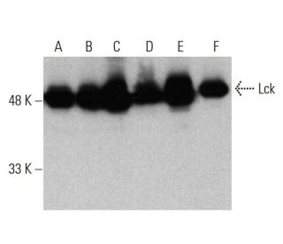
Lck 항체 (3A5): sc-433
- Lck 항체 3A5 는 마우스 monoclonal IgG2b κ Lck 항체, 313간행물에 인용, 이며 200 µg/ml으로 제공합니다.
- mouse origin의 Lck에서 내부에 위치한 1-225 아미노산을 항원으로 사용하였습니다.
- Lck 항체 (3A5)는 WB, IP, IF, IHC(P) and FCM으로 mouse, rat and human유래의 Lck p56 를 감지하는 데에 추천한다.
- 항-Lck 항체(3A5)는 IP용 agarose와 결합되어 이용 가능하며, WB, IHC(P), ELISA용 HRP와 결합되어 이용 가능하며, IF, IHC(P), FCM용 phycoerythrin 또는 FITC와 결합되어 이용 가능합니다.
- WB (RGB), IF, IHC(P) 와FCM, RGB fluorescent imaging systems, such as iBright™ FL1000, FluorChem™, Typhoon, Azure and other comparable systems에 사용가능한 Alexa Fluor® 488, Alexa Fluor® 546, Alexa Fluor® 594 or Alexa Fluor® 647결합제품도 있습니다.
- WB (NIR), IF와FCM,Near-Infrared (NIR) detection systems, such as LI-COR®Odyssey®, iBright™ FL1000, FluorChem™, Typhoon, Azure and other comparable systems에 사용가능한 Alexa Fluor® 680 or Alexa Fluor® 790 결합제품도 있습니다.
- 2b BP-HRP">m-IgG2b BP-HRP 및 m-IgGκ BP-HRP는 Lck 항체 (3A5) WB 및 IHC(P) 애플리케이션용입니다. 이 시약은 이제 Lck 항체 (3A5)와 함께 번들로 제공됩니다(아래 주문 정보 참조).

빠른 링크
더보기
Lck 항체(3A5)는 웨스턴 블롯팅(WB), 면역침전법(IP), 면역형광법(IF), 면역조직화학, 유세포분석법(FCM)을 통해 쥐, 쥐, 인간에서 유래된 Lck 단백질을 검출하는 쥐 단일클론 IgG2b 카파 경쇄 항체입니다. Lck (3A5) 항체는 비접합 형태와 아가로스, 과산화효소(HRP), 피코에리트린(PE), 플루오레세인 이소티오시아네이트(FITC), 다중 알렉사 플루오르® 접합체를 포함한 다양한 접합체 형태로 제공됩니다. Src 티로신 키나아제 계열의 일원인 Lck 단백질은 T 세포 신호 전달, 특히 T 세포 항원 수용체(TCR)의 활성화에 중요한 역할을 합니다. 이 활성화는 T 세포 증식, 분화, 사이토카인 생산으로 이어지는 하류 신호 경로를 촉발하기 때문에 면역 반응에 필수적입니다. Lck 단백질은 주로 T 세포의 원형질막에 위치하며, 이 곳에서 TCR 복합체와 결합하여 수용체 결합 시 신호를 효과적으로 전달할 수 있도록 합니다. 인간 Lck 유전자는 염색체 1p35.1에 위치해 있으며, 면역 반응 조절에 필수적인 509개의 아미노산으로 구성된 단백질을 암호화합니다. Lck의 기능과 국소화를 이해함으로써 연구자들은 T세포 생물학에서 Lck의 중요성과 면역학적 장애에 대한 잠재적 의미를 더 잘 이해할 수 있습니다.
Alexa Fluor®는 미국 오리건주 Molecular Probes Inc.의 상표입니다.
LI-COR®와 Odyssey®는 LI-COR Biosciences의 등록 상표입니다.
Lck 항체 (3A5) 참고문헌:
- Src 계열 키나아제 Lck 및 Fyn에 의해 매개되는 차별적 T세포 항원 수용체 신호. | Denny, MF., et al. 2000. Mol Cell Biol. 20: 1426-35. PMID: 10648627
- Src 계열의 키나아제: 구조와 기능. | Tatosyan, AG. and Mizenina, OA. 2000. Biochemistry (Mosc). 65: 49-58. PMID: 10702640
- 백혈구에서 Src 키나아제 매개 신호 전달. | Korade-Mirnics, Z. and Corey, SJ. 2000. J Leukoc Biol. 68: 603-13. PMID: 11073097
- Src 키나아제의 활성화와 기능에 대한 선택적 엿보기. | Bjorge, JD., et al. 2000. Oncogene. 19: 5620-35. PMID: 11114743
- SRC 계열 키나아제는 엔도텔린에 의한 상피 Na+ 채널 억제를 매개합니다. | Gilmore, ES., et al. 2001. J Biol Chem. 276: 42610-7. PMID: 11560932
- 아스파라긴은 LCK 신호를 강화하여 CD8+ T세포 활성화와 항암 반응을 촉진합니다. | Wu, J., et al. 2021. Nat Cell Biol. 23: 75-86. PMID: 33420490
- LAG3는 TCR-CD3 복합체와 결합하여 공동 수용체-Lck 해리를 유도함으로써 신호를 억제합니다. | Guy, C., et al. 2022. Nat Immunol. 23: 757-767. PMID: 35437325
- LCK 억제는 YAP 활동을 하향 조절하고, 환자 유래 담관암종 모델에서 치료 효과가 있습니다. | Conboy, CB., et al. 2023. J Hepatol. 78: 142-152. PMID: 36162702
- LCK가 아닌 FYN 키나아제 신호를 통한 CD28-CAR-T 세포 활성화는 치료 효과를 향상시킵니다. | Wu, L., et al. 2023. Cell Rep Med. 4: 100917. PMID: 36696897
- 부분적인 인간 LCK 결함은 장 염증을 동반한 T세포 면역결핍을 유발합니다. | Lui, VG., et al. 2024. J Exp Med. 221: PMID: 37962568
- 인간 원시 종양 유전자의 유전적 조직. | Sakaguchi, AY. 1983. Prog Clin Biol Res. 119: 93-103. PMID: 6191340
- Src 키나제 기능에 대한 인사이트: 구조적 비교. | Williams, JC., et al. 1998. Trends Biochem Sci. 23: 179-84. PMID: 9612082
주문정보
| 제품명 | 카탈로그 번호 | 단위 | 가격 | 수량 | 관심품목 | |
Lck 항체 (3A5) | sc-433 | 200 µg/ml | $322.00 | |||
Lck (3A5): m-IgGκ BP-HRP 번들 | sc-520408 | 200 µg Ab, 40 µg BP | $361.00 | |||
Lck (3A5): m-IgG2b BP-HRP 번들 | sc-548825 | 200 µg Ab; 10 µg BP | $361.00 | |||
Lck 항체 (3A5) AC | sc-433 AC | 500 µg/ml, 25% agarose | $424.00 | |||
Lck 항체 (3A5) HRP | sc-433 HRP | 200 µg/ml | $322.00 | |||
Lck 항체 (3A5) FITC | sc-433 FITC | 200 µg/ml | $336.00 | |||
Lck 항체 (3A5) PE | sc-433 PE | 200 µg/ml | $349.00 | |||
Lck 항체 (3A5) Alexa Fluor® 488 | sc-433 AF488 | 200 µg/ml | $364.00 | |||
Lck 항체 (3A5) Alexa Fluor® 546 | sc-433 AF546 | 200 µg/ml | $364.00 | |||
Lck 항체 (3A5) Alexa Fluor® 594 | sc-433 AF594 | 200 µg/ml | $364.00 | |||
Lck 항체 (3A5) Alexa Fluor® 647 | sc-433 AF647 | 200 µg/ml | $364.00 | |||
Lck 항체 (3A5) Alexa Fluor® 680 | sc-433 AF680 | 200 µg/ml | $364.00 | |||
Lck 항체 (3A5) Alexa Fluor® 790 | sc-433 AF790 | 200 µg/ml | $364.00 |