")
GFRα-1 Antibody (E-11): sc-271546
- GFRα-1 Antibody (E-11) is a mouse monoclonal IgG1 κ GFRα-1 antibody, cited in 33 publications, provided at 200 µg/ml
- raised against amino acids 368-437 of GFRα-1 of human origin
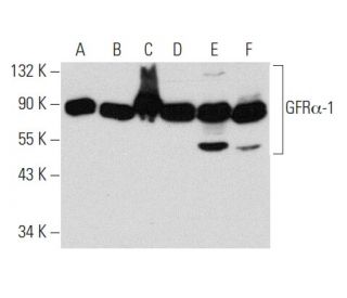
- GFR alpha-1 Antibody (E-11) is recommended for detection of GFRα-1 of mouse, rat and human origin by WB, IP, IF, IHC(P) and ELISA
- Anti-GFR alpha-1 Antibody (E-11) is available conjugated to agarose for IP; HRP for WB, IHC(P) and ELISA; and to either phycoerythrin or FITC for IF, IHC(P) and FCM
- also available conjugated to Alexa Fluor® 488, Alexa Fluor® 546, Alexa Fluor® 594 or Alexa Fluor® 647 for WB (RGB), IF, IHC(P) and FCM, and for use with RGB fluorescent imaging systems, such as iBright™ FL1000, FluorChem™, Typhoon, Azure and other comparable systems
- also available conjugated to Alexa Fluor® 680 or Alexa Fluor® 790 for WB (NIR), IF and FCM; for use with Near-Infrared (NIR) detection systems, such as LI-COR®Odyssey®, iBright™ FL1000, FluorChem™, Typhoon, Azure and other comparable systems
- m-IgG Fc BP-HRP, m-IgG1 BP-HRP and m-IgGκ BP-HRP are the preferred secondary detection reagents for GFRα-1 Antibody (E-11) for WB and IHC(P) applications. These reagents are now offered in bundles with GFRα-1 Antibody (E-11) (see ordering information below).

GFRα-1 Antibody (E-11) is a mouse monoclonal IgG1 kappa light chain antibody that detects GFRα-1 protein of mouse, rat, and human origin by western blotting (WB), immunoprecipitation (IP), immunofluorescence (IF), immunohistochemistry with paraffin-embedded sections (IHCP), and enzyme-linked immunosorbent assay (ELISA). GFRα-1 (E-11) antibody is available in both non-conjugated and various conjugated forms, including agarose, horseradish peroxidase (HRP), phycoerythrin (PE), fluorescein isothiocyanate (FITC), and multiple Alexa Fluor® conjugates. GFRα-1 plays a crucial role in neuronal survival and differentiation, primarily functioning as a receptor for glial cell line-derived neurotrophic factor (GDNF) and neurturin (NTN), which are essential for maintaining both central and peripheral neurons. GFRα-1 is located on the cell membrane, where a glycosyl-phosphoinositol linkage enables interaction with ligands and subsequent activation of tyrosine kinase Ret, leading to downstream signaling pathways that promote neuronal health. GFRα-1′s significance in neurotrophic signaling highlights potential therapeutic applications in neurodegenerative diseases, making anti-GFRα-1 antibody (E-11) an invaluable tool for studying neuronal function and survival mechanisms.
Alexa Fluor® is a trademark of Molecular Probes Inc., OR., USA
LI-COR® and Odyssey® are registered trademarks of LI-COR Biosciences
GFRα-1 Antibody (E-11) References:
- GFRalpha-1 mRNA in dopaminergic and nondopaminergic neurons in the substantia nigra and ventral tegmental area. | Sarabi, A., et al. 2001. J Comp Neurol. 441: 106-17. PMID: 11745638
- Evidence for a ligand-specific signaling through GFRalpha-1, but not GFRalpha-2, in the absence of Ret. | Pezeshki, G., et al. 2001. J Neurosci Res. 66: 390-5. PMID: 11746356
- XIB4035, a novel nonpeptidyl small molecule agonist for GFRalpha-1. | Tokugawa, K., et al. 2003. Neurochem Int. 42: 81-6. PMID: 12441171
- Induction of GDNF and GFRα-1 Following AAV1-Rheb(S16H) Administration in the Hippocampus in vivo. | Yun, D., et al. 2020. Exp Neurobiol. 29: 164-175. PMID: 32408406
- GFRα-1 is a reliable marker of bovine gonocytes/undifferentiated spermatogonia: A mini-review. | Zhang, XM. 2021. Anat Histol Embryol. 50: 13-14. PMID: 32761645
- Aberrantly expressed GFRα-1/RET in patients with lacrimal adenoid cystic carcinoma is associated with high recurrence risk: a retrospective study of 51 LACC cases. | Liu, L., et al. 2021. Cancer Biol Med. 18: 199-205. PMID: 33628594
- GDNF: a glial cell line-derived neurotrophic factor for midbrain dopaminergic neurons. | Lin, LF., et al. 1993. Science. 260: 1130-2. PMID: 8493557
- Characterization of a multicomponent receptor for GDNF. | Treanor, JJ., et al. 1996. Nature. 382: 80-3. PMID: 8657309
- GDNF-induced activation of the ret protein tyrosine kinase is mediated by GDNFR-alpha, a novel receptor for GDNF. | Jing, S., et al. 1996. Cell. 85: 1113-24. PMID: 8674117
- Neurturin, a relative of glial-cell-line-derived neurotrophic factor. | Kotzbauer, PT., et al. 1996. Nature. 384: 467-70. PMID: 8945474
- TrnR2, a novel receptor that mediates neurturin and GDNF signaling through Ret. | Baloh, RH., et al. 1997. Neuron. 18: 793-802. PMID: 9182803
- Expression and regulation of GFRalpha3, a glial cell line-derived neurotrophic factor family receptor. | Naveilhan, P., et al. 1998. Proc Natl Acad Sci U S A. 95: 1295-300. PMID: 9448325
Ordering Information
| Product Name | Catalog # | UNIT | Price | Qty | FAVORITES | |
GFRα-1 Antibody (E-11) | sc-271546 | 200 µg/ml | $322.00 | |||
GFRα-1 Antibody (E-11): m-IgG Fc BP-HRP Bundle | sc-529207 | 200 µg Ab; 10 µg BP | $361.00 | |||
GFRα-1 Antibody (E-11): m-IgGκ BP-HRP Bundle | sc-521975 | 200 µg Ab, 40 µg BP | $361.00 | |||
GFRα-1 Antibody (E-11): m-IgG1 BP-HRP Bundle | sc-543385 | 200 µg Ab; 20 µg BP | $361.00 | |||
GFRα-1 Antibody (E-11) AC | sc-271546 AC | 500 µg/ml, 25% agarose | $424.00 | |||
GFRα-1 Antibody (E-11) HRP | sc-271546 HRP | 200 µg/ml | $322.00 | |||
GFRα-1 Antibody (E-11) FITC | sc-271546 FITC | 200 µg/ml | $336.00 | |||
GFRα-1 Antibody (E-11) PE | sc-271546 PE | 200 µg/ml | $349.00 | |||
GFRα-1 Antibody (E-11) Alexa Fluor® 488 | sc-271546 AF488 | 200 µg/ml | $364.00 | |||
GFRα-1 Antibody (E-11) Alexa Fluor® 546 | sc-271546 AF546 | 200 µg/ml | $364.00 | |||
GFRα-1 Antibody (E-11) Alexa Fluor® 594 | sc-271546 AF594 | 200 µg/ml | $364.00 | |||
GFRα-1 Antibody (E-11) Alexa Fluor® 647 | sc-271546 AF647 | 200 µg/ml | $364.00 | |||
GFRα-1 Antibody (E-11) Alexa Fluor® 680 | sc-271546 AF680 | 200 µg/ml | $364.00 | |||
GFRα-1 Antibody (E-11) Alexa Fluor® 790 | sc-271546 AF790 | 200 µg/ml | $364.00 |