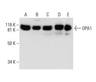
Date published: 2025-9-7

00800 4573 8000

SCBT Portrait Logo
Seach Input

Anticorpo OPA1 (D-9): sc-393296

4.0(22)
Scrivi una recensioneFai una domanda

Schede Tecniche
  • OPA1 Antibody D-9 è un monoclonale di topo IgG1 κ OPA1 antibody, citato in 62 pubblicazioni, fornito in 200 µg/ml
  • generato contro gli amminoacidi 647-780 mappati nella regione interna di OPA1 di origine human
  • OPA1 Antibody (D-9) é raccomandato per il rilevamento di OPA1 di origine mouse, rat e human in WB, IP, IF e ELISA; anche reattivo con ulteriori specie, incluse e equine, canine, bovine and porcine
  • OPA1 Antibody (D-9) é disponibile coniugato con agarose per IP; HRP per WB, IHC(P) ed ELISA; sia con phycoerythrin o FITC per IF, IHC(P) e FCM
  • disponibile anche coniugato con Alexa Fluor® 488, Alexa Fluor® 546; Alexa Fluor® 594 o Alexa Fluor® 647 per IF, IHC (P) e FCM
  • disponibile anche coniugato con Alexa Fluor® 680 o Alexa Fluor® 790 per WB (NIR), IF e FCM
  • Contattaci per ricevere un FREE 10 µg sample di OPA1 (D-9): sc-393296.
  • m-IgG Fc BP-HRP e m-IgGκ BP-HRP sono i reagenti secondari preferiti per OPA1 Anticorpo (D-9) per applicazioni WB. Questi reagenti sono ora offerti in bundle con OPA1 Anticorpo (D-9)(vedere le informazioni per l'ordine sotto).

LINK RAPIDI

VEDI ANCHE...

L'anticorpo OPA1 (D-9) è un anticorpo monoclonale di topo IgG1 κ OPA1 (designato anche anticorpo Dynamin-Like 120 KDa Protein Mitocondrial, anticorpo Dynamin-Like Guanosine Triphosphatase, anticorpo Optic Atrophy 1 (Autosomal Dominant), anticorpo Dynamin-Like GTPase mitocondriale, anticorpo Optic Atrophy Protein 1, anticorpo MGM1, anticorpo NPG, anticorpo NTG o anticorpo BERHS) che rileva la proteina OPA1 di origine murina, ratta e umana mediante WB, IP, IF ed ELISA. L'anticorpo OPA1 (D-9) è disponibile sia nella forma di anticorpo anti-OPA1 non coniugato, sia in molteplici forme coniugate di anticorpo anti-OPA1, tra cui agarosio, HRP, PE, FITC e molteplici coniugati Alexa Fluor®. OPA1 (proteina del gene dell'atrofia ottica 1), appartiene alla famiglia delle dinamine. Il gene che codifica OPA1 si localizza in 3q29, ha come target i mitocondri ed è coinvolto nella biogenesi mitocondriale. I difetti di OPA1 sono una causa dell'atrofia ottica di tipo 1. OPA1 è espresso principalmente nella retina, ma può essere espresso anche nel cervello, nel testicolo, nel cuore e nei muscoli scheletrici.

Solo per ricerca in vitro. Non destinato ad uso Diagnostico o Terapeutico.

Alexa Fluor® è un marchio registrato di Molecular Probes Inc., OR., USA

LI-COR® e Odyssey® sono marchi registrati di LI-COR Biosciences

Anticorpo OPA1 (D-9) Riferimenti:

  1. Il gene nucleare OPA1, che codifica una proteina mitocondriale legata alla dinamina, è mutato nell'atrofia ottica dominante.  |  Delettre, C., et al. 2000. Nat Genet. 26: 207-10. PMID: 11017079
  2. Spettro, frequenza e penetranza delle mutazioni di OPA1 nell'atrofia ottica dominante.  |  Toomes, C., et al. 2001. Hum Mol Genet. 10: 1369-78. PMID: 11440989
  3. Spettro di mutazioni e varianti di splicing nel gene OPA1.  |  Delettre, C., et al. 2001. Hum Genet. 109: 584-91. PMID: 11810270
  4. Sublocalizzazione differenziale delle isoforme della proteina legata alla dinamina OPA1 nei mitocondri.  |  Satoh, M., et al. 2003. Biochem Biophys Res Commun. 300: 482-93. PMID: 12504110
  5. Ruolo dei mediatori di fissione e fusione mitocondriale Fis1, Drp1 e Opa1 nell'apoptosi nei mammiferi.  |  Lee, YJ., et al. 2004. Mol Biol Cell. 15: 5001-11. PMID: 15356267
  6. OPA1 richiede la mitofusina 1 per promuovere la fusione mitocondriale.  |  Cipolat, S., et al. 2004. Proc Natl Acad Sci U S A. 101: 15927-32. PMID: 15509649
  7. OPA1, associato all'atrofia ottica autosomica dominante, è ampiamente espresso nel cervello umano.  |  Bette, S., et al. 2005. Acta Neuropathol. 109: 393-9. PMID: 15700187
  8. Raffinamento del locus dominante dell'atrofia ottica (OPA1) in un intervallo di 1,4 cm sul cromosoma 3q28-3q29, all'interno di un contig YAC di 3 Mb.  |  Jonasdottir, A., et al. 1997. Hum Genet. 99: 115-20. PMID: 9003507

Informazioni ordini

Nome del prodottoCodice del prodottoUNITÀPrezzoQuantitàPreferiti

OPA1 Anticorpo (D-9)

sc-393296
200 µg/ml
$316.00

OPA1 (D-9): m-IgG Fc BP-HRP Bundle

sc-530422
200 µg Ab; 10 µg BP
$354.00

OPA1 (D-9): m-IgGκ BP-HRP Bundle

sc-523893
200 µg Ab, 40 µg BP
$354.00

OPA1 Anticorpo (D-9) AC

sc-393296 AC
500 µg/ml, 25% agarose
$416.00

OPA1 Anticorpo (D-9) HRP

sc-393296 HRP
200 µg/ml
$316.00

OPA1 Anticorpo (D-9) FITC

sc-393296 FITC
200 µg/ml
$330.00

OPA1 Anticorpo (D-9) PE

sc-393296 PE
200 µg/ml
$343.00

OPA1 Anticorpo (D-9) Alexa Fluor® 488

sc-393296 AF488
200 µg/ml
$357.00

OPA1 Anticorpo (D-9) Alexa Fluor® 546

sc-393296 AF546
200 µg/ml
$357.00

OPA1 Anticorpo (D-9) Alexa Fluor® 594

sc-393296 AF594
200 µg/ml
$357.00

OPA1 Anticorpo (D-9) Alexa Fluor® 647

sc-393296 AF647
200 µg/ml
$357.00

OPA1 Anticorpo (D-9) Alexa Fluor® 680

sc-393296 AF680
200 µg/ml
$357.00

OPA1 Anticorpo (D-9) Alexa Fluor® 790

sc-393296 AF790
200 µg/ml
$357.00

I am currently looking for antibodies for OPA1 for immunofluorescence. I would like to ask if it is possible to get a trial sample to try? We have OPA1 ko cells so if it works, I could upload some photos and protocols to share. Many thanks in advance!

Richiesta da: Xiaotong
Please contact our Technical Service Department for more information on how to receive a free 10 ug sample of this monoclonal antibody.
Risposta da: Technical Support
Data di pubblicazione: 2020-07-24

As I am unhappy with the current OPA1-antibody we are using (other manufacturer), I am looking to buy a new one. Would it be possible to get a free sample of this antibody for testing?

Richiesta da: Christiaan
Thank you for your question. Please contact Technical Service for information on how to receive a free 10 ug sample of this monoclonal antibody.
Risposta da: Tech Service
Data di pubblicazione: 2020-02-06

8% separation gel, first antibody 1:100, 4℃,overnight. second antibody 1:1000,room temperature, 1 hour. But I think the band (130-250kDa) is not right.

Richiesta da: Anonimo
Thank you for your question. Please contact your local distributor or Technical Service directly for assistance regarding your WB results, we are happy to discuss your results with you.
Risposta da: Tech Service
Data di pubblicazione: 2019-04-26

what buffer does the opa1 antibody work best in? I tried probing a blot of mouse heart tissue with opa1 at 1:1000 in 5%milk/ TBST and the blot is there is a lot of background. I checked the publications cited and they lack the buffer details

Richiesta da: Dr C
Thank you for your question. The optimal blocking buffer and antibody diluent depends on which type of Western blot detection method you are using. If you are using an HRP conjugated secondary for chemiluminescent detection, then we recommend Blotto, or 5% nonfat milk in TBST (1X TBST + 0.05% Tween-20) for blocking, and for incubating both primary and secondary antibodies. If you are using a fluorescent or NIR (Near-Infrared) detection method, for optimal results, use low fluorescence PVDF membranes, such as Immobilon®-FL PVDF: sc-516541, and UltraCruz® Blocking Reagent: sc-516214 for all blocking, primary antibody and secondary antibody incubations.
Risposta da: Technical Support
Data di pubblicazione: 2022-04-27

I would like to know your OPA1 antibody (D9): sc-393296 could use with immunofluorescence? How long to be shipped to Korea, the volume and the price to my university in Korea. Thank you very much!

Richiesta da: Trang
Thank you for your question. We have not yet tested this mouse monoclonal antibody, OPA1 (D-9): sc-393296, in immunofluorescent staining applications. However, based on the antibody's performance in other applications, we do recommend and warranty the antibody for IF, or immunofluorescent staining of fresh or frozen tissues or cells, fixed in methanol or acetone. Please contact one of our authorized distributors in Korea to inquire about shipping times and to receive a quote to ship to a University in Korea. A link is provided below to search for our distributors. https://www.scbt.com/scbt/resources/distributors For a limited time, we are offering a free 10 µg sample of this antibody. Please contact us if you would like to request a sample to test in your application. Please call our Asian Support office toll-free at 800-820-8626, email asia@scbio.cn, or contact us by live chat.
Risposta da: Technical Support
Data di pubblicazione: 2017-05-25

Can OPA1 Antibody (D-9): sc-393296 be used for detection of OPA1 of chicken origin?

Richiesta da: jenni
Thank you for your inquiry. Please contact our Technical Service Department and we would be happy to perform sequence analysis and review our data to answer any questions you may have about additional species reactivity. We are available by phone, e-mail or chat.
Risposta da: Technical Support 3
Data di pubblicazione: 2017-01-09
  • y_2025, m_8, d_31, h_8CST
  • bvseo_bulk, prod_bvqa, vn_bulk_3.0.42
  • cp_1, bvpage1
  • co_hasquestionsanswers, tq_6
  • loc_it_IT, sid_393296, prod, sort_[SortEntry(order=LAST_APPROVED_ANSWER_SUBMISSION_TIME, direction=DESCENDING)]
  • clientName_scbt
  • bvseo_sdk, java_sdk, bvseo-4.0.0
  • CLOUD, getContent, 195ms
  • QUESTIONS, PRODUCT
Rated 1 di 5 di da it does not worksMaybe it works in lysates from tissue but does not works in cell lysates, not even at 1:100.
Data di pubblicazione: 2023-04-24
Rated 1 di 5 di da very poor quality.Purchased this antibody for studying mitochondrial dynamics, very faint signal and a huge background noise, I used 1/100 dilution. after 4 times trying with different gels and samples, I gave up.
Data di pubblicazione: 2021-04-01
Rated 4 di 5 di da goodSingle extremely weak band detected with muscle cells, but strong single in muscle tissue.
Data di pubblicazione: 2020-02-06
Rated 4 di 5 di da Good Inner mitochondrial membrane protein markerCan be used in 1:500 dilution for western blotting to look at mouse mitochondrial extracts.
Data di pubblicazione: 2019-10-05
Rated 5 di 5 di da good signalI loaded total homogenate from mouse brain 5-10-20-30 ug, good signal with 20 and 30 ug. I recommend this antibody for WB.
Data di pubblicazione: 2019-01-28
Rated 3 di 5 di da Not quite strong band1:200 diluted for wb of rat protein,the band is not quite clear
Data di pubblicazione: 2018-06-12
Rated 3 di 5 di da WBTested in SHSY-5Y human neuroblastoma cells (~15-20ug/lane), dilution 1:1000 in 3% milk in TBST. Got a single clear band at expected size after very long exposure.
Data di pubblicazione: 2018-05-15
Rated 4 di 5 di da Great featuresI bought it a month ago, it works well as I recent run in WB.
Data di pubblicazione: 2017-10-30
  • y_2025, m_8, d_31, h_8
  • bvseo_bulk, prod_bvrr, vn_bulk_3.0.42
  • cp_1, bvpage1
  • co_hasreviews, tv_0, tr_22
  • loc_it_IT, sid_393296, prod, sort_[SortEntry(order=SUBMISSION_TIME, direction=DESCENDING)]
  • clientName_scbt
  • bvseo_sdk, java_sdk, bvseo-4.0.0
  • CLOUD, getReviews, 16ms
  • REVIEWS, PRODUCT
Anticorpo OPA1 (D-9) valutazione 4.0 di 5 di 22.
  • y_2025, m_8, d_31, h_8
  • bvseo_bulk, prod_bvrr, vn_bulk_3.0.42
  • cp_1, bvpage1
  • co_hasreviews, tv_0, tr_22
  • loc_it_IT, sid_393296, prod, sort_[SortEntry(order=SUBMISSION_TIME, direction=DESCENDING)]
  • clientName_scbt
  • bvseo_sdk, java_sdk, bvseo-4.0.0
  • CLOUD, getAggregateRating, 179ms
  • REVIEWS, PRODUCT