")
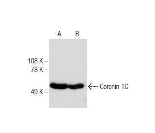
: sc-376919. Analisi Western blot dell'espressione di Coronina 1C in estratto nucleare di HeLa (A) e lisato cellulare intero di T24 (B).")
: sc-376919. Colorazione con immunoperossidasi di tessuto epatico umano fissato in formalina e incluso in paraffina che mostra la colorazione della membrana delle cellule del dotto biliare e la colorazione citoplasmatica e della membrana dei sinusoidi epatici.")
: sc-376919. Analisi in Western blot dell'espressione della Coronina 1C in estratti nucleari di HeLa (A) e NIH/3T3 (B), lisati cellulari interi di Neuro-2A (C) ed estratti di tessuto di cervello di topo (D) e ippocampo di ratto (E).")
Anticorpo Coronin 1C (D-9): sc-376919
- Coronin 1C Antibody D-9 è un monoclonale di topo IgM κ Coronin 1C antibody, citato in 3 pubblicazioni, fornito in 200 µg/ml
- generato contro gli amminoacidi 390-474 mappati al C-terminus di Coronin 1C di origine human
- raccomandato per il rilevamento di Coronin 1C di origine mouse, rat e human in WB, IP, IF, IHC(P) e ELISA
- m-IgGκ BP-HRP è il reagente secondario di rilevazione preferito per l'anticorpo Coronin 1C (D-9) per applicazioni WB e IHC(P). Questo reagente è ora offerto in combinazione con l'anticorpo Coronin 1C (D-9)(vedere le informazioni per l'ordine sotto). Per ulteriori coniugati m-IgGκ BP, consultare l'elenco completo delle proteine leganti le IgG di topo.

LINK RAPIDI
L'anticorpo Coronin 1C (D-9) è un anticorpo monoclonale di topo IgM κ Coronin 1C (designato anche come anticorpo CORO1C o anticorpo Coronin 3) che rileva la proteina Coronin 1C di origine murina, ratta e umana mediante WB, IP, IF, IHC(P) ed ELISA. L'anticorpo Coronina 1C (D-9) è disponibile come anticorpo anti-Coronina 1C non coniugato. Le coronine sono una famiglia di proteine contenenti ripetizioni WD che legano l'actina e che si localizzano nelle aree sottomembrana e regolano la motilità cellulare e il riarrangiamento del citoscheletro. La coronina 1A (CORO1A, CLIPINA, CLABP, TACO, p57) può formare complessi omotrimerici mediati da coiled coil che influenzano la formazione precoce dei fagosomi. La fosforilazione dipendente da PKC della Coronina 1B (CORO1B) alla serina 2 regola la dinamica del bordo d'attacco e la motilità cellulare nei fibroblasti attraverso le interazioni con il complesso Arp2/3. La coronina 1C (CORO1C, Coronina 3, HCRNN4) è abbondante nelle cellule Neuro-2a in fase di differenziamento, nelle cellule PC-12 e negli oligodendrociti primari, dove si ritiene che influenzi la morfogenesi e la migrazione dei neuroni. La coronina 2A (CORO2A, CLIPINB, IR10, WDR2) è un componente del complesso N-CoR (nuclear receptor corepressor) di circa 1,5-2 megadalton, composto da 10-12 proteine, che recluta le HDAC per generare cromatina repressiva. La coronina 7 (CORO7, CRN7) si localizza sulla membrana del Golgi e influenza l'organizzazione dei compartimenti di membrana intracellulare e il traffico vescicolare. La coronina 2B (CORO2B, CLIPINC) e la coronina 6 (CORO6) sono simili ad altri membri di questa famiglia, poiché possiedono un motivo basico N-terminale conservato e 3-10 ripetizioni WD raggruppate in uno o due domini centrali.
Alexa Fluor® è un marchio registrato di Molecular Probes Inc., OR., USA
LI-COR® e Odyssey® sono marchi registrati di LI-COR Biosciences
Anticorpo Coronin 1C (D-9) Riferimenti:
- La coronina si localizza ai bordi di attacco ed è coinvolta nella diffusione cellulare e nell'estensione del lamellipodio nelle cellule dei vertebrati. | Mishima, M. and Nishida, E. 1999. J Cell Sci. 112 (Pt 17): 2833-42. PMID: 10444378
- L'oligomerizzazione, l'interazione con la F-actina e l'associazione alla membrana della coronina 3, ubiquitaria nei mammiferi, sono mediate dal suo terminale carbossilico. | Spoerl, Z., et al. 2002. J Biol Chem. 277: 48858-67. PMID: 12377779
- Purificazione e caratterizzazione funzionale del complesso N-CoR umano: i ruoli di HDAC3, TBL1 e TBLR1. | Yoon, HG., et al. 2003. EMBO J. 22: 1336-46. PMID: 12628926
- La coronina 7, l'omologo della POD-1 dei mammiferi, si localizza nell'apparato del Golgi. | Rybakin, V., et al. 2004. FEBS Lett. 573: 161-7. PMID: 15327992
- Associazione della membrana plasmatica dei leucociti con il citoscheletro di actina attraverso molecole trimeriche di coronina 1 mediate da coiled coil. | Gatfield, J., et al. 2005. Mol Biol Cell. 16: 2786-98. PMID: 15800061
- La coronina 3 e il suo ruolo nella morfogenesi cerebrale murina. | Hasse, A., et al. 2005. Eur J Neurosci. 21: 1155-68. PMID: 15813925
- La funzione di Coronin-1 è necessaria per la formazione del fagosoma. | Yan, M., et al. 2005. Mol Biol Cell. 16: 3077-87. PMID: 15829569
- Le proteine coronine come regolatori multifunzionali del citoscheletro e del traffico di membrana. | Rybakin, V. and Clemen, CS. 2005. Bioessays. 27: 625-32. PMID: 15892111
- La fosforilazione della coronina 1B da parte della protein chinasi C regola l'interazione con Arp2/3 e la motilità cellulare. | Cai, L., et al. 2005. J Biol Chem. 280: 31913-23. PMID: 16027158
- La coronina-1C è un nuovo biomarcatore per la progressione invasiva del carcinoma epatocellulare identificato mediante analisi proteomica e validazione clinica. | Wu, L., et al. 2010. J Exp Clin Cancer Res. 29: 17. PMID: 20181269
- La coronina 1C promuove l'invasività del carcinoma mammario triplo negativo attraverso la regolazione del traffico MT1-MMP e della funzione invadopodia. | Castagnino, A., et al. 2018. Oncogene. 37: 6425-6441. PMID: 30065298
- La coronina 1C, regolata da microRNA multipli, facilita l'aggressività delle cellule tumorali nell'adenocarcinoma duttale pancreatico. | Fukuda, K., et al. 2023. Genes (Basel). 14: PMID: 37239355
Informazioni ordini
| Nome del prodotto | Codice del prodotto | UNITÀ | Prezzo | Quantità | Preferiti | |
Coronin 1C Anticorpo (D-9) | sc-376919 | 200 µg/ml | $322.00 | |||
Coronin 1C (D-9): m-IgGκ BP-HRP Bundle | sc-552447 | 200 µg Ab; 40 µg BP | $361.00 |