")
COPE Antikörper (A-4): sc-133195
- COPE Antikörper A-4 ist ein Maus monoklonales IgG2a κ COPE Antikörper, verwendet in 1 wissenschaftlichen Veröffentlichungen, in einer Menge von 200 µg/ml
- gezogen gegen die Aminosäuresequenz 111-190 von COPE aus der Spezies human
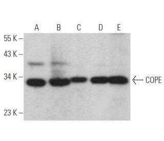
- COPE Antikörper (A-4) ist empfohlen für die Detektion von COPE aus der Spezies mouse, rat und human per WB, IP, IF, IHC(P) und ELISA
- Anti-COPE Antikörper (A-4) ist erhältlich als Konjugat mit Agarose für IP; HRP für WB, IHC(P) und ELISA; und entweder mit Phycoerythrin oder FITC für IF, IHC(P) und FCM
- auch erhältlich als Konjugat mit Alexa Fluor® 488, Alexa Fluor® 546, Alexa Fluor® 594 oder Alexa Fluor® 647 für IF, IHC(P) und FCM
- auch erhältlich als Konjugat mit Alexa Fluor® 680 oder Alexa Fluor® 790 für WB (NIR), IF und FCM
- m-IgG Fc BP-HRP, 2a BP-HRP">m-IgG2a BP-HRP und m-IgGκ BP-HRP sind die bevorzugten sekundären Nachweisreagenzien für COPE Antikörper (A-4) für WB- und IHC(P)-Anwendungen. Diese Reagenzien werden jetzt in Bündeln mit COPE Antikörper (A-4) angeboten(siehe Bestellinformationen unten).

Der COPE-Antikörper (A-4) ist ein monoklonaler IgG2a-Antikörper der leichten Kette Kappa der Maus, der das COPE-Protein von Mäusen, Ratten und Menschen durch Western Blot (WB), Immunopräzipitation (IP), Immunfluoreszenz (IF), Immunhistochemie und Enzyme-linked Immunosorbent Assay (ELISA) nachweist. Der anti-COPE-Antikörper (A-4) ist sowohl in nicht konjugierter als auch in verschiedenen konjugierten Formen erhältlich, darunter Agarose, Meerrettichperoxidase (HRP), Phycoerythrin (PE), Fluoresceinisothiocyanat (FITC) und mehrere Alexa Fluor®-Konjugate. Das COPE-Protein spielt eine entscheidende Rolle beim Membran- und Vesikeltransport innerhalb des frühen Sekretionswegs, insbesondere bei der Vermittlung des retrograden Transports vom Golgi-Apparat zurück zum endoplasmatischen Retikulum sowie beim Intra-Golgi-Transport durch mit Nicht-Clathrin-Coat-Protein I beschichtete Vesikel. Der heptamere Coatomer-Komplex, der zytosolische Vorläufer der COPI-Hülle, besteht aus zwei Subkomplexen: Der erste umfasst die β-, γ-, δ- und ζ-COP-Untereinheiten, während der zweite die α-COP-, COPP- und ε-COP-Untereinheiten umfasst. Das Verständnis der Struktur und Funktion des COPE-Proteins ist von entscheidender Bedeutung, da das COPE-Protein für die Aufrechterhaltung der zellulären Homöostase und die Gewährleistung einer ordnungsgemäßen Proteinsortierung und -beförderung unerlässlich ist, die für verschiedene zelluläre Prozesse und die allgemeine Zellfunktion von entscheidender Bedeutung sind.
Alexa Fluor® ist ein Markenzeichen von Molecular Probes Inc., OR., USA
LI-COR® und Odyssey® sind Markenzeichen von LI-COR Biosciences
COPE Antikörper (A-4) Literaturhinweise:
- Die Ausrichtung von Proteinen auf das endoplasmatische Retikulum durch Dilysin-Signale beinhaltet nicht nur eine direkte Retention, sondern auch eine Abrufung. | Andersson, H., et al. 1999. J Biol Chem. 274: 15080-4. PMID: 10329713
- Der epitheliale Na(+)/H(+)-Austauscher, NHE3, wird über einen Clathrin-vermittelten Weg internalisiert. | Chow, CW., et al. 1999. J Biol Chem. 274: 37551-8. PMID: 10608808
- Beeinträchtigte Proteasomfunktion rettet die Thermosensitivität von Hefezellen, denen die Coatomer-Untereinheit epsilon-COP fehlt. | Kimata, Y., et al. 2000. J Biol Chem. 275: 10655-60. PMID: 10744762
- In vitro Montage und Demontage von Coatomer. | Lowe, M. and Kreis, TE. 1995. J Biol Chem. 270: 31364-71. PMID: 8537409
- Hemmung der Endosomenfunktion in CHO-Zellen, die einen temperaturempfindlichen Defekt in der Coatomer (COPI)-Komponente epsilon-COP tragen. | Daro, E., et al. 1997. J Cell Biol. 139: 1747-59. PMID: 9412469
- epsilon-COP ist ein struktureller Bestandteil des Coatomers, der die Funktion hat, alpha-COP zu stabilisieren. | Duden, R., et al. 1998. EMBO J. 17: 985-95. PMID: 9463377
- Eine einzige Bindungsstelle für Dilysin-Retrieval-Motive und p23 innerhalb der Gamma-Untereinheit von Coatomer. | Harter, C. and Wieland, FT. 1998. Proc Natl Acad Sci U S A. 95: 11649-54. PMID: 9751720
Bestellinformation
| Produkt | Katalog # | EINHEIT | Preis | ANZAHL | Favoriten | |
COPE Antikörper (A-4) | sc-133195 | 200 µg/ml | $322.00 | |||
COPE (A-4): m-IgG Fc BP-HRP Bundle | sc-528766 | 200 µg Ab; 10 µg BP | $361.00 | |||
COPE (A-4): m-IgGκ BP-HRP Bundle | sc-521271 | 200 µg Ab, 40 µg BP | $361.00 | |||
COPE (A-4): m-IgG2a BP-HRP Bundle | sc-547164 | 200 µg Ab; 10 µg BP | $361.00 | |||
COPE Antikörper (A-4) AC | sc-133195 AC | 500 µg/ml, 25% agarose | $424.00 | |||
COPE Antikörper (A-4) HRP | sc-133195 HRP | 200 µg/ml | $322.00 | |||
COPE Antikörper (A-4) FITC | sc-133195 FITC | 200 µg/ml | $336.00 | |||
COPE Antikörper (A-4) PE | sc-133195 PE | 200 µg/ml | $349.00 | |||
COPE Antikörper (A-4) Alexa Fluor® 488 | sc-133195 AF488 | 200 µg/ml | $364.00 | |||
COPE Antikörper (A-4) Alexa Fluor® 546 | sc-133195 AF546 | 200 µg/ml | $364.00 | |||
COPE Antikörper (A-4) Alexa Fluor® 594 | sc-133195 AF594 | 200 µg/ml | $364.00 | |||
COPE Antikörper (A-4) Alexa Fluor® 647 | sc-133195 AF647 | 200 µg/ml | $364.00 | |||
COPE Antikörper (A-4) Alexa Fluor® 680 | sc-133195 AF680 | 200 µg/ml | $364.00 | |||
COPE Antikörper (A-4) Alexa Fluor® 790 | sc-133195 AF790 | 200 µg/ml | $364.00 |