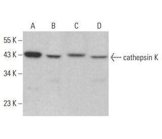
Date published: 2025-9-7

00800 4573 8000

SCBT Portrait Logo
Seach Input

Anticorpo cathepsin K (E-7): sc-48353

5.0(20)
Scrivi una recensioneFai una domanda

Schede Tecniche
  • cathepsin K Antibody E-7 è un monoclonale di topo IgG1 κ cathepsin K antibody, citato in 165 pubblicazioni, fornito in 200 µg/ml
  • contrari agli amminoacidi 191-240 mappati in una regione interna del dominio cathepsin K di origine human
  • raccomandato per il rilevamento di cathepsin K di origine mouse, rat e human in WB, IP, IF, IHC(P) e ELISA
  • disponibile coniugato a HRP per WB, IHC(P) ed ELISA; e ad altri phycoerythrina o FITC, Alexa Fluor® 488 o Alexa Fluor® 647 per IF, IHC(P) e FCM
  • Contattaci per ricevere un FREE 10 µg sample di cathepsin K (E-7): sc-48353.
  • m-IgG Fc BP-HRP e m-IgGκ BP-HRP sono i reagenti di rivelazione secondaria preferiti per cathepsin K anticorpo (E-7) per le applicazioni WB and IHC(P). Questi reagenti sono ora offerti in un pacchetto con cathepsin K Anticorpo (E-7) (vedi informazioni per l'ordine sotto).

LINK RAPIDI

VEDI ANCHE...

L'anticorpo anti-cathepsina K (E-7) è un anticorpo monoclonale di topo IgG1 a catena leggera kappa che rileva la proteina cathepsina K di origine murina, di ratto e umana mediante western blotting (WB), immunoprecipitazione (IP), immunofluorescenza (IF), immunoistochimica con sezioni incluse in paraffina (IHCP) e saggio di immunoassorbimento enzimatico (ELISA). L'anticorpo anti-catepsina K (E-7) è disponibile sia in forma non coniugata che in varie forme coniugate, tra cui agarosio, perossidasi di rafano (HRP), ficoeritrina (PE), isotiocianato di fluoresceina (FITC) e molteplici coniugati Alexa Fluor®. La catepsina K, membro della famiglia delle proteasi cisteiniche, svolge un ruolo cruciale nel riassorbimento e nel rimodellamento osseo, rendendo la catepsina K essenziale per mantenere la salute e l'omeostasi ossea. Questo enzima è espresso prevalentemente negli osteoclasti, cellule responsabili della degradazione ossea, e l'attività della catepsina K è fondamentale per il normale turnover osseo fisiologico. La disregolazione della catepsina K è stata implicata in vari disturbi ossei, tra cui l'osteoporosi e la malattia di Paget, evidenziando l'importanza della catepsina K come bersaglio terapeutico. La capacità dell'anticorpo anti-cathepsina K (E-7) di rilevare più specie migliora la ricerca e le applicazioni cliniche, consentendo una comprensione più approfondita del metabolismo osseo e delle malattie correlate.

Solo per ricerca in vitro. Non destinato ad uso Diagnostico o Terapeutico.

Alexa Fluor® è un marchio registrato di Molecular Probes Inc., OR., USA

LI-COR® e Odyssey® sono marchi registrati di LI-COR Biosciences

Anticorpo cathepsin K (E-7) Riferimenti:

  1. Clonazione molecolare ed espressione della catepsina S dei macrofagi alveolari umani, una proteasi cisteinica elastinolitica.  |  Shi, GP., et al. 1992. J Biol Chem. 267: 7258-62. PMID: 1373132
  2. Organizzazione molecolare del gene della catepsina D umana.  |  Redecker, B., et al. 1991. DNA Cell Biol. 10: 423-31. PMID: 2069717
  3. Inibitori della catepsina K per l'osteoporosi: biologia, potenziale utilità clinica e lezioni apprese.  |  Drake, MT., et al. 2017. Endocr Rev. 38: 325-350. PMID: 28651365
  4. Le cellule progenitrici di derivazione tendinea che esprimono catepsina K attivano la segnalazione di Hedgehog per guidare l'ossificazione eterotopica.  |  Feng, H., et al. 2020. J Clin Invest. 130: 6354-6365. PMID: 32853181
  5. Clonazione molecolare e sequenziamento del cDNA della catepsina L di ratto.  |  Ishidoh, K., et al. 1987. FEBS Lett. 223: 69-73. PMID: 3666143
  6. Clonazione molecolare e sequenziamento del cDNA della catepsina H di ratto. Omologia nelle regioni pro-peptidiche delle proteinasi cisteiniche.  |  Ishidoh, K., et al. 1987. FEBS Lett. 226: 33-7. PMID: 3691815
  7. L'inibizione della catepsina K induce la destabilizzazione di Raptor e la disfunzione mitocondriale attraverso la segnalazione mediata dall'asse Syk/SHP2/Src/OTUB1.  |  Seo, SU., et al. 2023. Cell Death Dis. 14: 366. PMID: 37330581
  8. Isolamento e sequenziamento di due cloni di cDNA codificanti la catepsina E della milza di ratto e analisi dell'attivazione della procatepsina E purificata.  |  Okamoto, K., et al. 1995. Arch Biochem Biophys. 322: 103-11. PMID: 7574663
  9. Clonazione molecolare della catepsina O umana, una nuova endoproteinasi e omologo della OC2 del coniglio.  |  Shi, GP., et al. 1995. FEBS Lett. 357: 129-34. PMID: 7805878
  10. La catepsina B, una proteasi cisteinica implicata nella progressione metastatica, è espressa anche durante la regressione della prostata e delle ghiandole mammarie del ratto.  |  Guenette, RS., et al. 1994. Eur J Biochem. 226: 311-21. PMID: 8001549
  11. Clonazione molecolare, localizzazione cromosomica ed espressione tessuto-specifica del gene della catepsina G murina.  |  Heusel, JW., et al. 1993. Blood. 81: 1614-23. PMID: 8453108
  12. Catepsina K del topo: clonazione del cDNA ed espressione predominante del gene negli osteoclasti e in alcuni condrociti ipertrofici durante lo sviluppo del topo.  |  Rantakokko, J., et al. 1996. FEBS Lett. 393: 307-13. PMID: 8814310

Informazioni ordini

Nome del prodottoCodice del prodottoUNITÀPrezzoQuantitàPreferiti

cathepsin K Anticorpo (E-7)

sc-48353
200 µg/ml
$316.00

cathepsin K (E-7): m-IgG Fc BP-HRP Bundle

sc-528526
200 µg Ab; 10 µg BP
$354.00

cathepsin K (E-7): m-IgGκ BP-HRP Bundle

sc-520922
200 µg Ab, 40 µg BP
$354.00

cathepsin K Anticorpo (E-7) Alexa Fluor® 488

sc-48353 AF488
200 µg/ml
$357.00

cathepsin K Anticorpo (E-7) Alexa Fluor® 647

sc-48353 AF647
200 µg/ml
$357.00

cathepsin K Anticorpo (E-7) FITC

sc-48353 FITC
200 µg/ml
$330.00

cathepsin K Anticorpo (E-7) HRP

sc-48353 HRP
200 µg/ml
$316.00

cathepsin K Anticorpo (E-7) PE

sc-48353 PE
200 µg/ml
$343.00

I cannot find an antibody to Cathepsin K that can be used in rabbit. There are Cathepsin K antibodies available raised in rabbit but that should give a very high back ground and may lead to false positives. Any suggestions?

Richiesta da: Doty
Thank you for your question, this antibody is not warrantied for rabbit, but it does have a high homology for rabbit. This would be what we'd recommend for your sample.
Risposta da: Technical Service 4
Data di pubblicazione: 2020-12-21

What concentration should this be used at for flow cytometry? Presumably this is after fix/perm?

Richiesta da: SB KCL
Thank you for your question. Please note that unconjugated cathepsin K Antibody (E-7): sc-48353 is not recommended for flow cytometry. You can find our standard protocol under this link: https://www.scbt.com/resources/protocols/flow-cytometry We recommend using 1 µg of fluorochrome-conjugated antibodies per tube with 1 million cells.
Risposta da: Technical Support Europe
Data di pubblicazione: 2020-10-21

For WB, is it possible to use secondary Fc-specific antibody to detect E-7 (sc-48353) antibody ?

Richiesta da: TomG
Thank you for your inquiry and your interest in this product. The cathepsin K Antibody (E-7): sc-48353 is a mouse monoclonal IgG3 containing a kappa light chain. You can certainly use a conventional Fc-specific anti-mouse secondary antibody for detection. However, we would rather recommend to use the directly conjugated formats of this antibody or our Mouse IgG Kappa Binding Proteins, e.g. m-IgGκ BP-HRP: sc-516102. Please contact our European Support Office in Germany directly for any further questions you may have. You can reach us by phone at: +49 6221 4503 0, by email at: europe@scbt.com or by live chat directly on our website, www.scbt.com We are happy to assist you.
Risposta da: Technical Support Europe
Data di pubblicazione: 2018-01-04

For IHC, which antigen retrieval method is recommended?

Richiesta da: rbrun
Thank you for your question. We recommend using sodium citrate buffer (pH 6.0) with heat treatment for antigen retrieval with this antibody, cathepsin K (E-7): sc-48353. A link to our recommended protocol is provided below as a reference. https://www.scbt.com/scbt/resources/protocols/immunoperoxidase-staining
Risposta da: Technical Support
Data di pubblicazione: 2024-12-03

For Western Blot, is it recommended to use denatured or non-denatured conditions with this antibody?

Richiesta da: AbPolly
Thank you for your question. We recommend this antibody for use in denatured Western Blot conditions. It has not been validated for use in non-denatured conditions. Please contact our Technical Service Department for further details or inquiries.
Risposta da: Technical Support 12
Data di pubblicazione: 2017-01-04
  • y_2025, m_8, d_28, h_5CST
  • bvseo_bulk, prod_bvqa, vn_bulk_3.0.42
  • cp_1, bvpage1
  • co_hasquestionsanswers, tq_5
  • loc_it_IT, sid_48353, prod, sort_[SortEntry(order=LAST_APPROVED_ANSWER_SUBMISSION_TIME, direction=DESCENDING)]
  • clientName_scbt
  • bvseo_sdk, java_sdk, bvseo-4.0.0
  • CLOUD, getContent, 108ms
  • QUESTIONS, PRODUCT
Rated 5 di 5 di da 非常好的一只抗体!在Western blot实验中表现出色一年多前购买了这只抗体,效果非常的不错。非常好的一只抗体!在Western blot实验中表现出色!
Data di pubblicazione: 2024-12-02
Rated 5 di 5 di da Very specific antibodyWe used these antibody to analyze active cathepsin K in mouse and human osteoclast. You can see our results in the following publication: Dey, G. et al, Development and Application of Reversible and Irreversible Covalent Probes for Human and Mouse Cathepsin‐K Activity Detection, Revealing Nuclear Activity. Advanced Science 2024. https://doi.org/10.1002/advs.202401518.
Data di pubblicazione: 2024-11-25
Rated 4 di 5 di da Cathepsin KDot Blot was perfomed to detect the Cathepsin K in the cell culture supernatants.
Data di pubblicazione: 2020-07-29
Rated 5 di 5 di da Great signal!Works well in 1:500 dilution to stain cathepsin K from mouse bone marrow-derived osteoclast samples.
Data di pubblicazione: 2019-03-22
Rated 5 di 5 di da Works well1:500 dilution, clear bands in mouse BMMs induced to osteoclast by RANKL
Data di pubblicazione: 2018-01-17
Rated 5 di 5 di da Excellent stainingI have received this sample and used it for staining of cat K in mice aortic tissue and cultured vascular smooth muscle cells. The staining was very clear and excellent.
Data di pubblicazione: 2017-10-27
Rated 5 di 5 di da great signalUsing 1/1000 diluted the antibody to work in WB, and there were bands in the mouse tissue, ~39kDa, which was a good result.
Data di pubblicazione: 2017-10-24
Rated 5 di 5 di da Worked very well!I tried IHC-P in mouse tissue (bone) and the results were very nice at 1:100.
Data di pubblicazione: 2017-09-22
  • y_2025, m_8, d_28, h_5
  • bvseo_bulk, prod_bvrr, vn_bulk_3.0.42
  • cp_1, bvpage1
  • co_hasreviews, tv_0, tr_20
  • loc_it_IT, sid_48353, prod, sort_[SortEntry(order=SUBMISSION_TIME, direction=DESCENDING)]
  • clientName_scbt
  • bvseo_sdk, java_sdk, bvseo-4.0.0
  • CLOUD, getReviews, 19ms
  • REVIEWS, PRODUCT
Anticorpo cathepsin K (E-7) valutazione 5.0 di 5 di 20.
  • y_2025, m_8, d_28, h_5
  • bvseo_bulk, prod_bvrr, vn_bulk_3.0.42
  • cp_1, bvpage1
  • co_hasreviews, tv_0, tr_20
  • loc_it_IT, sid_48353, prod, sort_[SortEntry(order=SUBMISSION_TIME, direction=DESCENDING)]
  • clientName_scbt
  • bvseo_sdk, java_sdk, bvseo-4.0.0
  • CLOUD, getAggregateRating, 109ms
  • REVIEWS, PRODUCT