")
cathepsin K Anticorpo (E-7): sc-48353
- cathepsin K Anticorpo E-7é um anticorpo monoclonal produzido em camundongo IgG1 κ cathepsin K anticorpo, citado em 165 publicações, fornecido em 200 µg/ml
- suscitadas contra os aminoácidos 191-240 mapeados numa região interna de cathepsin K de origem human
- recomendado para a detecção de cathepsin K de mouse, rat e human origem em
- disponível conjugado a HRP para WB, IHC(P) e ELISA; e tanto para a ficoeritrina ou FITC, Alexa Fluor® 488 ou Alexa Fluor® 647 para IF, IHC(P) e FCM
- O m-IgG Fc BP-HRP e o m-IgGκ BP-HRP são os reagentes de deteção secundários preferidos para o anticorpo cathepsin K (E-7) para aplicações WB e IHC(P). Estes reagentes são agora disponibilizados em conjuntos com o anticorpo cathepsin K (E-7)(ver informações de encomenda abaixo).

LINKS RÁPIDOS
VEJA TAMBÉM
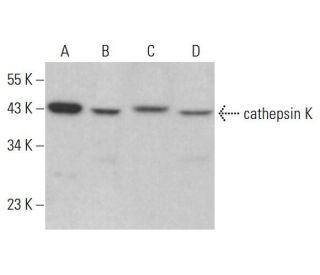
O anticorpo contra a catepsina K (E-7) é um anticorpo monoclonal IgG1 de cadeia leve kappa de ratinho que detecta a proteína catepsina K de origem camundongo, ratazana e humana por western blotting (WB), imunoprecipitação (IP), imunofluorescência (IF), imuno-histoquímica com secções incluídas em parafina (IHCP) e ensaio de imunoabsorção enzimática (ELISA). O anticorpo anti-catepsina K (E-7) está disponível em formas não conjugadas e em várias formas conjugadas, incluindo agarose, peroxidase de rábano (HRP), ficoeritrina (PE), isotiocianato de fluoresceína (FITC) e vários conjugados Alexa Fluor®. A catepsina K, um membro da família da cisteína protease, desempenha um papel crucial na reabsorção e remodelação óssea, tornando a catepsina K essencial para manter a saúde e a homeostasia ósseas. Esta enzima é predominantemente expressa nos osteoclastos, células responsáveis pela degradação óssea, e a atividade da catepsina K é vital para a renovação fisiológica normal do osso. A desregulação da catepsina K tem sido implicada em várias doenças ósseas, incluindo a osteoporose e a doença de Paget, o que realça a importância da catepsina K como alvo terapêutico. A capacidade do anticorpo anti-catepsina K (E-7) para detetar em várias espécies melhora a investigação e as aplicações clínicas, permitindo uma compreensão mais profunda do metabolismo ósseo e das doenças relacionadas.
Alexa Fluor® é uma marca comercial da Molecular Probes Inc., OR., EUA
LI-COR® e Odyssey® são marcas registadas da LI-COR Biosciences
Referencias do cathepsin K Anticorpo (E-7):
- Clonagem molecular e expressão da catepsina S de macrófagos alveolares humanos, uma cisteína protease elastinolítica. | Shi, GP., et al. 1992. J Biol Chem. 267: 7258-62. PMID: 1373132
- Organização molecular do gene da catepsina D humana. | Redecker, B., et al. 1991. DNA Cell Biol. 10: 423-31. PMID: 2069717
- Inibidores da catepsina K para a osteoporose: Biologia, utilidade clínica potencial e lições aprendidas. | Drake, MT., et al. 2017. Endocr Rev. 38: 325-350. PMID: 28651365
- Células progenitoras derivadas do tendão que expressam catepsina K activam a sinalização Hedgehog para impulsionar a ossificação heterotópica. | Feng, H., et al. 2020. J Clin Invest. 130: 6354-6365. PMID: 32853181
- Clonagem molecular e sequenciação do cDNA da catepsina L do rato. | Ishidoh, K., et al. 1987. FEBS Lett. 223: 69-73. PMID: 3666143
- Clonagem molecular e sequenciação do cDNA da catepsina H do rato. Homologia nas regiões pro-peptídicas das cisteíno-proteinases. | Ishidoh, K., et al. 1987. FEBS Lett. 226: 33-7. PMID: 3691815
- A inibição da catepsina K induz a desestabilização do Raptor e a disfunção mitocondrial através da sinalização mediada pelo eixo Syk/SHP2/Src/OTUB1. | Seo, SU., et al. 2023. Cell Death Dis. 14: 366. PMID: 37330581
- Isolamento e sequenciação de dois clones de cDNA que codificam a catepsina E do baço de rato e análise da ativação da procatepsina E purificada. | Okamoto, K., et al. 1995. Arch Biochem Biophys. 322: 103-11. PMID: 7574663
- Clonagem molecular da catepsina O humana, uma nova endoproteinase e homóloga da OC2 do coelho. | Shi, GP., et al. 1995. FEBS Lett. 357: 129-34. PMID: 7805878
- A catepsina B, uma cisteína protease implicada na progressão metastática, também é expressa durante a regressão da próstata e das glândulas mamárias do rato. | Guenette, RS., et al. 1994. Eur J Biochem. 226: 311-21. PMID: 8001549
- Clonagem molecular, localização cromossómica e expressão tecido-específica do gene da catepsina G murina. | Heusel, JW., et al. 1993. Blood. 81: 1614-23. PMID: 8453108
- Catepsina K do rato: clonagem do cDNA e expressão predominante do gene nos osteoclastos e em alguns condrócitos hipertrofiados durante o desenvolvimento do rato. | Rantakokko, J., et al. 1996. FEBS Lett. 393: 307-13. PMID: 8814310
Informacoes sobre ordens
| Nome do Produto | Numero de Catalogo | UNID | Preco | Qde | FAVORITOS | |
cathepsin K Anticorpo (E-7) | sc-48353 | 200 µg/ml | $322.00 | |||
Pacote do cathepsin K (E-7): m-IgG Fc BP-HRP | sc-528526 | 200 µg Ab; 10 µg BP | $361.00 | |||
Pacote do cathepsin K (E-7): m-IgGκ BP-HRP | sc-520922 | 200 µg Ab, 40 µg BP | $361.00 | |||
cathepsin K Anticorpo (E-7) Alexa Fluor® 488 | sc-48353 AF488 | 200 µg/ml | $364.00 | |||
cathepsin K Anticorpo (E-7) Alexa Fluor® 647 | sc-48353 AF647 | 200 µg/ml | $364.00 | |||
cathepsin K Anticorpo (E-7) FITC | sc-48353 FITC | 200 µg/ml | $336.00 | |||
cathepsin K Anticorpo (E-7) HRP | sc-48353 HRP | 200 µg/ml | $322.00 | |||
cathepsin K Anticorpo (E-7) PE | sc-48353 PE | 200 µg/ml | $349.00 |