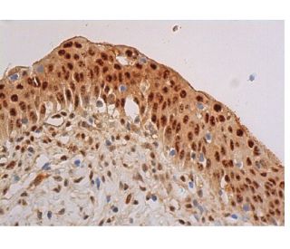
Date published: 2025-9-7

00800 4573 8000

SCBT Portrait Logo
Seach Input

p-Akt1/2/3 Anticuerpo (B-12): sc-377556

4.2(10)
Escribir una reseñaHacer una pregunta

Fichas Técnicas
  • p-Akt1/2/3 Anticuerpo (B-12) es un monoclonal de ratón IgM κ, ver las 38 publicaciones, proporcionado como 200 µg/ml
  • específica para un epítopo cartográfico entre los aminoácidos Thr 450 Thr 450 de Akt1 de human origen
  • recomendado para detectarThr 450 phosphorylated Akt1 and Thr 451 correspondingly phosphorylated Akt2 and Thr 447 correspondingly phosphorylated Akt3 de mouse, rat y human origen, mediante WB, IP, IF, IHC(P) y ELISA; también reactivo con otras especies, incluyendo anyd equine, bovine and porcine
  • Contacte con nosotros para recibir GRATIS 10 µg de muestra de p-Akt1/2/3 (B-12): sc-377556.
  • Actualmente, aún no hemos completado la identificación de los reactivos de detección secundaria preferidos para p-Akt1/2/3 Anticuerpo (B-12). Este trabajo está en progreso.

ENLACES RÁPIDOS

VER TAMBIÉN ....

El anticuerpo p-Akt1/2/3 (B-12) es un anticuerpo monoclonal de ratón IgM κ que detecta la Akt1 fosforilada en Treonina 450 y la Akt2 fosforilada en Treonina 451, así como la Akt3 fosforilada en Treonina 447, de origen humano, de rata y de ratón, mediante las técnicas de WB, IP, IF, IHC(P) y ELISA. El anticuerpo p-Akt1/2/3 (B-12) está disponible en su forma no conjugada. La familia de quinasas serina/treonina Akt contiene varios miembros, incluyendo Akt1 (también designada como PKB o RacPK), Akt2 (también designada como PKBβ o RacPK-β) y Akt3 (también designada como PKBγ o protooncogén viral de timo 3), los cuales muestran homología de secuencia con las familias de quinasa de proteína A y C y son codificados por el protooncogén c-Akt. Todos los miembros de la familia Akt tienen un dominio de homología de pleckstrin. Akt1 y Akt2 son activados por la estimulación de PDGF. Esta activación depende de los residuos de tirosina 740 y 751 de PDGFR-β, los cuales se unen a la subunidad del complejo de fosfatidilinositol 3-quinasa (PI 3-quinasa). La activación de Akt1 por insulina o factor de crecimiento insulínico-1 (IGF-1) resulta en la fosforilación tanto de Treonina 308 como de Serina 473. Las proteínas Akt se fosforilan y activan en células estimuladas por insulina/IGF-1 por una quinasa(s) aguas arriba, y la activación de Akt1 y Akt2 es inhibida por el inhibidor de PI quinasa wortmannin. En conjunto, estos datos sugieren fuertemente que la proteína señala aguas abajo de las quinasas PI. Akt3 es fosforilada en un residuo de serina en respuesta a la insulina. Sin embargo, la activación de Akt3 por insulina es inhibida por la activación previa de la proteína quinasa C a través de un mecanismo que no requiere la presencia del dominio PH. Akt3 se expresa en fibroblastos 3T3-L1, adipocitos y músculo esquelético y puede estar involucrada en varios procesos biológicos, incluyendo la diferenciación de adipocitos y músculo, síntesis de glucógeno, captación de glucosa, apoptosis y proliferación celular.

Para uso exclusivo en Investigación No está diseñado para para Diagnóstico ó Terapia.

Información sobre pedidos

Nombre del productoNúmero de catálogoUNIDADPrecioCANTIDADFavoritos

p-Akt1/2/3 Anticuerpo (B-12)

sc-377556
200 µg/ml
$316.00

p-Akt1/2/3 (B-12) péptido neutralizante

sc-377556 P
100 µg/0.5 ml
$68.00

I am using B-12 antibody (pAKT), is it igM second antibody with HRP to be used for detection? or will it work with igGk second antibody?

Preguntado por: Chai 123
Thank you for your question. p-Akt1/2/3 Antibody (B-12) is a mouse monoclonal IgM and we do recommend using an isotype specific secondary anti-mouse IgM for detection. Alternatively, you could also use our mouse IgG kappa binding protein (m-IgGκ BP) conjugated to Horseradish Peroxidase (HRP), m-IgGκ BP-HRP: sc-516102. This secondary antibody binds specifically to the kappa light chain of mouse IgG, and is also reactive with the kappa light chain of mouse IgM. A link to the product information is provided below for your reference. https://www.scbt.com/scbt/product/m-igg-kappa-bp-hrp
Respondida por: Technical Support
Fecha de publicación: 2017-06-29

What application is the blocking peptide sc-377556 P appropriate for?

Preguntado por: DefinitelyNotMatt
Thank you for your question. The blocking peptide is intended for use as a negative control, by pre-adsorbing the mouse monoclonal antibody against the antigen. For full protocol details, please contact our Technical Services Department or view our online protocol here: https://www.scbt.com/scbt/resources/protocols/peptide-neutralization
Respondida por: Technical Support
Fecha de publicación: 2017-02-24
  • y_2025, m_8, d_29, h_8CST
  • bvseo_bulk, prod_bvqa, vn_bulk_3.0.42
  • cp_1, bvpage1
  • co_hasquestionsanswers, tq_2
  • loc_es_ES, sid_377556, prod, sort_[SortEntry(order=LAST_APPROVED_ANSWER_SUBMISSION_TIME, direction=DESCENDING)]
  • clientName_scbt
  • bvseo_sdk, java_sdk, bvseo-4.0.0
  • CLOUD, getContent, 109ms
  • QUESTIONS, PRODUCT
Rated 4 de 5 por de Showed specific bandsGood antibody showed specific band with 1:500 dilution.
Fecha de publicación: 2017-10-20
Rated 4 de 5 por de Not very sureSample was mixed with 5X Reducing Loading Buffer and Denature the proteins by heating the sample to 95°C. Load the sample onto 3% Stacking Gel and 10% Separation Gel and run the gel under standard conditions. Transfer the proteins to a PVDF membrane using semi-dry or wet transfer methods. Block the membrane with 3% w/v skim milk powder in TBST. Incubate for 2 hr with agitation. Dilute the primary antibody in WB Anti-body Diluent (1:200) to the recommended dilution. Incubate overnight at 4°C, and rinse the blot in TBST four times for 5 min each at room temperature. Dilute the HRP labeled goat anti-mouse secondary antibody at the Second Ab dilute (1/5000). Incubate 1hr at room temperature, and rinse the blot in four times for 5 min each at room temperature. Perform ECL Plus detection. The result shows below: Not bands, hoverever, were showed in the membrane. That may because our lab used chicken sample, the result testified its antibody also cannot apply in chicken sample.
Fecha de publicación: 2017-10-18
Rated 4 de 5 por de Good antibodyWorked well with western blotting at 1:500 dilution.
Fecha de publicación: 2017-09-27
Rated 4 de 5 por de Gute Ergebnisse in der IHC(P) auf Herz/RatteMit Standardprotokoll ohne AG Demaskierung ON/4°C, mouse IgGkappa BP. Gute Ergebnisse in der IHC(P) auf Herz/Ratte.
Fecha de publicación: 2017-08-15
Rated 5 de 5 por de great antibodyThe result is good , although there is background .the band is clear.
Fecha de publicación: 2017-07-24
Rated 3 de 5 por de Nice productThere were some slight background but nice product.
Fecha de publicación: 2017-05-08
Rated 5 de 5 por de Worked Great!I was given a sample of this and it has worked great!
Fecha de publicación: 2017-03-27
Rated 3 de 5 por de Suitable antibody for Western blottingThis monoclonal antibody was independently tested by Kinexus Bioinformatics Corporation and is recommended for Western blotting. There was medium detection of ~ 60 kDa Akt in overnight serum-starved human A431 cells treated for 5 minutes with EGF on Western blots, and no significant cross-reactivities on these blots with any off-target proteins, except for weak-medium ~80, 135 and 150 kDa proteins.
Fecha de publicación: 2017-03-14
  • y_2025, m_8, d_29, h_8
  • bvseo_bulk, prod_bvrr, vn_bulk_3.0.42
  • cp_1, bvpage1
  • co_hasreviews, tv_0, tr_10
  • loc_es_ES, sid_377556, prod, sort_[SortEntry(order=SUBMISSION_TIME, direction=DESCENDING)]
  • clientName_scbt
  • bvseo_sdk, java_sdk, bvseo-4.0.0
  • CLOUD, getReviews, 16ms
  • REVIEWS, PRODUCT
p-Akt1/2/3 Anticuerpo (B-12) clasificado 4.2 de 5 por 10.
  • y_2025, m_8, d_29, h_8
  • bvseo_bulk, prod_bvrr, vn_bulk_3.0.42
  • cp_1, bvpage1
  • co_hasreviews, tv_0, tr_10
  • loc_es_ES, sid_377556, prod, sort_[SortEntry(order=SUBMISSION_TIME, direction=DESCENDING)]
  • clientName_scbt
  • bvseo_sdk, java_sdk, bvseo-4.0.0
  • CLOUD, getAggregateRating, 117ms
  • REVIEWS, PRODUCT