")
Tns3 Antikörper (C-2): sc-376367
- Tns3 Antikörper C-2 ist ein Maus monoklonales IgG2a κ Tns3 Antikörper, verwendet in 2 wissenschaftlichen Veröffentlichungen, in einer Menge von 200 µg/ml
- gezogen gegen die Aminosäuresequenz 721-1020 lokalisiert innerhalb einer internen Region von Tns3 aus der Spezies human
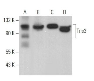
- Tns3 Antikörper (C-2) ist empfohlen für die Detektion von Tns3 aus der Spezies mouse, rat und human per WB, IP, IF, IHC(P) und ELISA
- Anti-Tns3 Antikörper (C-2) ist erhältlich als Konjugat mit Agarose für IP; HRP für WB, IHC(P) und ELISA; und entweder mit Phycoerythrin oder FITC für IF, IHC(P) und FCM
- auch erhältlich als Konjugat mit Alexa Fluor® 488, Alexa Fluor® 546, Alexa Fluor® 594 oder Alexa Fluor® 647 für IF, IHC(P) und FCM
- auch erhältlich als Konjugat mit Alexa Fluor® 680 oder Alexa Fluor® 790 für WB (NIR), IF und FCM
- m-IgG Fc BP-HRP, 2a BP-HRP">m-IgG2a BP-HRP und m-IgGκ BP-HRP sind die bevorzugten sekundären Nachweisreagenzien für Tns3 Antikörper (C-2) für WB- und IHC(P)-Anwendungen. Diese Reagenzien werden jetzt in Bündeln mit Tns3 Antikörper (C-2) angeboten(siehe Bestellinformationen unten).

Direktverknüpfungen
Der Tns3-Antikörper (C-2) ist ein monoklonaler Maus IgG2a κ Tns3-Antikörper (auch als Tns3-Antikörper bezeichnet), der das Tns3-Protein von Maus-, Ratte- und menschlicher Herkunft mittels WB, IP, IF, IHC (P) und ELISA nachweist. Der Tns3-Antikörper (C-2) ist sowohl in nicht konjugierter Form als auch in mehreren konjugierten Formen des Tns3-Antikörpers erhältlich, darunter Agarose, HRP, PE, FITC und mehrere Alexa Fluor®-Konjugate. Die Tensin (Tns)-Familie von Proteinen ist an der Erhaltung der Zellstruktur beteiligt, indem sie Aktinfilamente über F-Aktin-Bindungs- und Kapselungsaktivitäten an der Fokaladhesion ankern. Tns-Proteine enthalten auch ein Src-Homology-2 (SH2)-Domäne und haben die Fähigkeit, phosphoryliert zu werden, was auf eine Rolle in Signaltransduktionskaskaden hinweist. Diese vielfältigen Eigenschaften deuten darauf hin, dass Tns-Proteine wichtige Verbindungen zwischen dem Zytoskelett und Signaltransduktionswegen sein können. Tns3, auch als TEM6 oder TENS1 bekannt, lokalisiert sich an der Fokaladhesion der Plasmamembran. Es wird hauptsächlich in Schilddrüse und Plazenta exprimiert, kann aber auch im Herzen, Leber, Gehirn, Prostata, Pankreas, Niere, Lunge, Skelettmuskel und weißen Blutzellen gefunden werden. Tns3 ist für ein korrektes Wachstum und eine korrekte Entwicklung unerlässlich, wie durch das Wachstumsretardierung und den Tod einer Reihe von Tns3-/-Mäusen nahegelegt wird.
Alexa Fluor® ist ein Markenzeichen von Molecular Probes Inc., OR., USA
LI-COR® und Odyssey® sind Markenzeichen von LI-COR Biosciences
Tns3 Antikörper (C-2) Literaturhinweise:
- Der epidermale Wachstumsfaktor moduliert die Tyrosinphosphorylierung eines neuen Mitglieds der Tensinfamilie, Tensin3. | Cui, Y., et al. 2004. Mol Cancer Res. 2: 225-32. PMID: 15140944
- Die Inaktivierung von Tensin3 bei Mäusen führt zu Wachstumsverzögerung und postnataler Letalität. | Chiang, MK., et al. 2005. Dev Biol. 279: 368-77. PMID: 15733665
- Tensin3 ist ein neues schilddrüsenspezifisches Gen. | Maeda, I., et al. 2006. J Mol Endocrinol. 36: R1-8. PMID: 16461921
- Tensin: ein potenzielles Bindeglied zwischen dem Zytoskelett und der Signaltransduktion. | Lo, SH., et al. 1994. Bioessays. 16: 817-23. PMID: 7840759
- Molekulare Klonierung, Expression und Kartierung der hochaffinen Aktin-Capping-Domäne des kardialen Tensins von Hühnern. | Chuang, JZ., et al. 1995. J Cell Biol. 128: 1095-109. PMID: 7896874
- Interaktionen von Tensin mit Aktin und Identifizierung seiner drei verschiedenen Aktin-Bindungsdomänen. | Lo, SH., et al. 1994. J Cell Biol. 125: 1067-75. PMID: 8195290
- Die Ausbreitung von Zellen auf extrazellulären Matrixproteinen induziert die Tyrosinphosphorylierung von Tensin. | Bockholt, SM. and Burridge, K. 1993. J Biol Chem. 268: 14565-7. PMID: 8325835
- Die N-terminalen Domänen von Tensin und Auxilin sind Phosphatase-Homologe. | Haynie, DT. and Ponting, CP. 1996. Protein Sci. 5: 2643-6. PMID: 8976573
Bestellinformation
| Produkt | Katalog # | EINHEIT | Preis | ANZAHL | Favoriten | |
Tns3 Antikörper (C-2) | sc-376367 | 200 µg/ml | $322.00 | |||
Tns3 (C-2): m-IgG Fc BP-HRP Bundle | sc-529859 | 200 µg Ab; 10 µg BP | $361.00 | |||
Tns3 (C-2): m-IgGκ BP-HRP Bundle | sc-522929 | 200 µg Ab, 40 µg BP | $361.00 | |||
Tns3 (C-2): m-IgG2a BP-HRP Bundle | sc-547392 | 200 µg Ab; 10 µg BP | $361.00 | |||
Tns3 Antikörper (C-2) AC | sc-376367 AC | 500 µg/ml, 25% agarose | $424.00 | |||
Tns3 Antikörper (C-2) HRP | sc-376367 HRP | 200 µg/ml | $322.00 | |||
Tns3 Antikörper (C-2) FITC | sc-376367 FITC | 200 µg/ml | $336.00 | |||
Tns3 Antikörper (C-2) PE | sc-376367 PE | 200 µg/ml | $349.00 | |||
Tns3 Antikörper (C-2) Alexa Fluor® 488 | sc-376367 AF488 | 200 µg/ml | $364.00 | |||
Tns3 Antikörper (C-2) Alexa Fluor® 546 | sc-376367 AF546 | 200 µg/ml | $364.00 | |||
Tns3 Antikörper (C-2) Alexa Fluor® 594 | sc-376367 AF594 | 200 µg/ml | $364.00 | |||
Tns3 Antikörper (C-2) Alexa Fluor® 647 | sc-376367 AF647 | 200 µg/ml | $364.00 | |||
Tns3 Antikörper (C-2) Alexa Fluor® 680 | sc-376367 AF680 | 200 µg/ml | $364.00 | |||
Tns3 Antikörper (C-2) Alexa Fluor® 790 | sc-376367 AF790 | 200 µg/ml | $364.00 |